

Microcystic serous cystadenoma of the pancreas causing biliary obstruction: a case report and review of the literature
- PMID: 38463732
- PMCID: PMC10924709
- DOI: 10.1093/jscr/rjae105
Microcystic serous cystadenoma of the pancreas causing biliary obstruction: a case report and review of the literature
Abstract
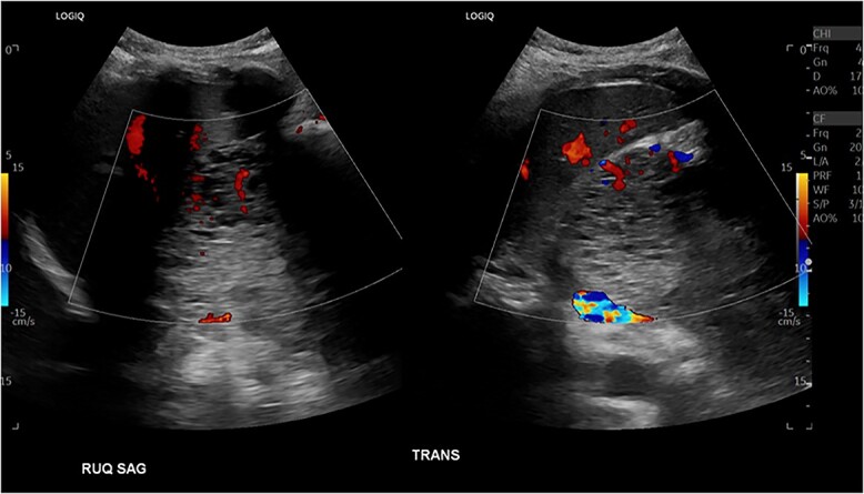
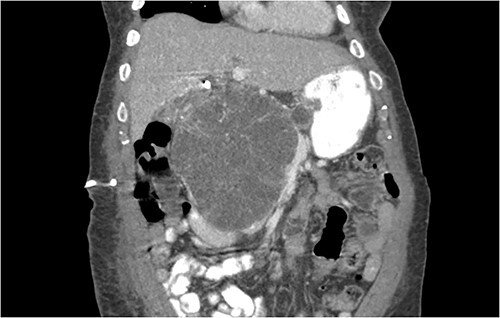
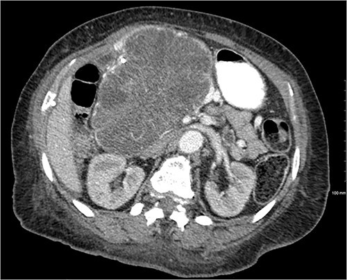
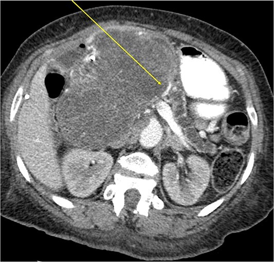
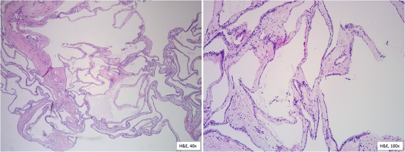
Cystic tumors account for 15% of pancreatic tumors. Of these, serous microcystic adenomas represent 1-2% of pancreatic exocrine neoplasms. While typically benign, a small percentage possess malignant potential. Given imaging improvements, serous cystadenomas are being identified more frequently. A 63-year-old female was admitted with complaints of jaundice and unintentional weight loss. Abdominal computed tomography scan showed a 16 cm obstructive pancreatic mass near the porta hepatis region. Endoscopic ultrasonography and fine needle aspiration biopsy indicated a large pancreatic head cystic mass favoring serous microcystadenoma causing biliary and some pyloric obstruction. Malignant potential could not be ruled out because of size and symptoms. A pylorus-preserving pancreaticoduodenectomy revealed a cystic tumor invading the pancreatic duct and adhering to the duodenum of the pancreatic head. Pathology confirmed a 15 cm benign pancreatic serous cystadenoma. Although most serous cystadenomas are benign, surgical resection was prudent given the size, symptoms, and adjacent organ involvement.
Keywords: Whipple surgery; biliary obstruction; microcystic serous cystadenoma.
Published by Oxford University Press and JSCR Publishing Ltd. © The Author(s) 2024.
Conflict of interest statement
None declared.
Figures
Similar articles
-
Serous cystadenoma of the pancreas associated with obstructive jaundice.J Gastroenterol. 2003;38(5):501-6. doi: 10.1007/s00535-002-1089-0. J Gastroenterol. 2003. PMID: 12768395 Review.
-
[Serous microcystic adenoma of the head of the pancreas causing an obstructive jaundice].Vojnosanit Pregl. 2008 Nov;65(11):839-42. doi: 10.2298/vsp0811839c. Vojnosanit Pregl. 2008. PMID: 19069716 Serbian.
-
Benign macrocystic serous cystadenoma of the pancreas.Indian J Pathol Microbiol. 2021 Jun;64(Supplement):S166-S168. doi: 10.4103/IJPM.IJPM_945_19. Indian J Pathol Microbiol. 2021. PMID: 34135161
-
Obstructive Jaundice as a Complication of Macrocystic Serous Cystadenoma of the Pancreas.Acta Med Indones. 2016 Apr;48(2):129-33. Acta Med Indones. 2016. PMID: 27550882
-
[A case of serous cystadenoma of the pancreas associated with obstructive jaundice during long-term follow up].Nihon Shokakibyo Gakkai Zasshi. 2011 Jun;108(6):962-8. Nihon Shokakibyo Gakkai Zasshi. 2011. PMID: 21646764 Review. Japanese.
References
Publication types
LinkOut - more resources
Full Text Sources
