. 2024 Feb 24;10(5):e27122.
doi: 10.1016/j.heliyon.2024.e27122. eCollection 2024 Mar 15.

Mitochondrial DNA levels in perfusate and bile during ex vivo normothermic machine correspond with donor liver quality

Affiliations

Mitochondrial DNA levels in perfusate and bile during ex vivo normothermic machine correspond with donor liver quality

Lauren P Westhaver et al. Heliyon. .

Abstract

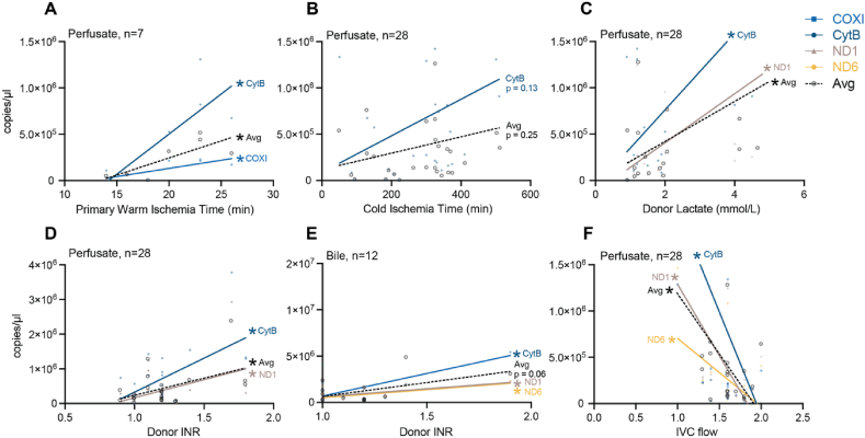
Ex vivo normothermic machine perfusion (NMP) preserves donor organs and permits real-time assessment of allograft health, but the most effective indicators of graft viability are uncertain. Mitochondrial DNA (mtDNA), released consequent to traumatic cell injury and death, including the ischemia-reperfusion injury inherent in transplantation, may meet the need for a biomarker in this context. We describe a real time PCR-based approach to assess cell-free mtDNA during NMP as a universal biomarker of allograft quality. Measured in the perfusate fluid of 29 livers, the quantity of mtDNA correlated with metrics of donor liver health including International Normalized Ratio (INR), lactate, and warm ischemia time, and inversely correlated with inferior vena cava (IVC) flow during perfusion. Our findings endorse mtDNA as a simple and rapidly measured feature that can inform donor liver health, opening the possibility to better assess livers acquired from extended criteria donors to improve organ supply.

Keywords: Extended criteria organ donation; Liver transplantation; Mitochondrial DAMPs; Mitochondrial DNA; Normothermic organ perfusion; Organ health assessment; Solid organ transplantation.


Conflict of interest statement

The authors declare that no conflict of interest exists.

Figures

Fig. 1
Fig. 1
Mitochondrial DNA is quantitated using gene target-specific synthetic oligonucleotide standard curves. SYBR green-based qPCR was performed with standard curves composed of serially diluted synthetic oligonucleotides corresponding to each primer-specific amplicon. (A) Representative qPCR amplification curves are shown for a two-fold serial dilution of oligonucleotides corresponding to the COX I-amplicon, from 107 to 76 copy numbers. (B) Standard curve formed by linear regression of Cq values plotted against log10 (copy number) of serially diluted COX I amplicon oligonucleotides (C) Representative qPCR melt curves and (D) melt peaks corresponding to the COX I amplification curves shown in A. (E) Gel electrophoresis of qPCR products corresponding to amplification curves for the COX I standard curve shown in A-D. (F) Comparison of mtDNA quantification from five healthy donor plasma samples using ddPCR, qPCR with a CytB-specific synthetic oligonucleotide standard curve (Synthetic oligo std qPCR). Each method of quantitation was performed over three independent qPCR experiments, each with a minimum of two technical replicates per sample. Bars/lines represent means ± SEM. (For interpretation of the references to colour in this figure legend, the reader is referred to the Web version of this article.)
Fig. 2
Fig. 2
MtDNA copy number in perfusate collected during ex vivo NMP of donor livers is consistent across gene targets. mtDNA copy number in perfusate from each donor liver was quantitated via qPCR with amplicon-specific standard curves for COX I, CytB, ND1, and ND6 at the final timepoint before transplantation. (A–F) Simple linear regressions were performed between pairs of mitochondrial gene targets at the last sequential NMP time point; correlations are reported as coefficient of determination (R2). Quantitation was performed over three independent qPCR experiments, each with a minimum of two technical replicates per sample.
Fig. 3
Fig. 3
Quantitation of mtDNA in perfusate and bile samples collected from donor livers during ex vivo NMP prior to transplant. MtDNA quantitation for perfusate samples collected at sequential NMP time points is shown for each donor liver and mtDNA gene target (A) COX I, (B) CytB, (C) ND1, or (D) ND6. Sequential samples for each patient (unique ID) are shown in the same vertical band; each data point from left to right represents earlier to later collection time points. qPCR products were subsequently assessed by agarose gel electrophoresis (Supplementary Fig. 1). (E) mtDNA levels for COX I, CytB, ND1, and ND6 in bile produced by a subset of donor livers during NMP. Bile samples were collected at the conclusion of NMP. Donor liver ID numbers correspond with ID numbers in A-D; bile production during NMP was not observed in every donor liver. Corresponding perfusate samples were unavailable for bile samples from donor liver ID numbers 30 to 35. qPCR products were subsequently assessed by agarose gel electrophoresis (Supplementary Fig. 2). Mean ± SEM is shown for each mtDNA gene target. Data represents three independent qPCR experiments, each with a minimum of two technical replicates per sample.
Fig. 4
Fig. 4
MtDNA levels in perfusate and bile collected during ex vivo NMP are significantly associated with donor and perfusion parameters. Simple linear regressions were performed for continuous variables and mtDNA copy number collected at the final time point during ex-vivo NMP. Those with statistically significant regressions, or near statistical significance, and the average of the four mtDNA copy number values are plotted (dotted line). For samples where matched clinical data was available (as indicated), regressions were performed between mtDNA level in perfusate and (A) primary warm ischemia time (WIT) incurred by donor livers prior to ex vivo NMP, (B) cold ischemia time (CIT) incurred by donor livers prior to ex vivo NMP, and (C) donor lactate measured prior to ex vivo NMP. (D, E) association between mtDNA levels in perfusate (D) and bile (E) with donor INR measured prior to ex vivo NMP. Donor INR data was not available for every donor liver that produced bile. (F) association between mtDNA levels in perfusate and IVC flow during NMP. Each data point represents the average of three independent qPCR experiments, each with a minimum of two technical replicates per sample.

References

    1. Dar W.A., Sullivan E., Bynon J.S., Eltzschig H., Ju C. Ischaemia reperfusion injury in liver transplantation: cellular and molecular mechanisms. Liver Int. 2019;39:788–801. doi: 10.1111/liv.14091. - DOI - PMC - PubMed
    1. Zhai Y., Petrowsky H., Hong J.C., Busuttil R.W., Kupiec-Weglinski J.W. Ischaemia-reperfusion injury in liver transplantation--from bench to bedside. Nat. Rev. Gastroenterol. Hepatol. 2013;10:79–89. doi: 10.1038/nrgastro.2012.225. - DOI - PMC - PubMed
    1. Czigany Z., Lurje I., Tolba R.H., Neumann U.P., Tacke F., Lurje G. Machine perfusion for liver transplantation in the era of marginal organs—new kids on the block. Liver Int. 2019;39:228–249. doi: 10.1111/liv.13946. - DOI - PubMed
    1. Pandya K., Sastry V., Panlilio M.T., Yip T.C.F., Salimi S., West C., Virtue S., Wells M., Crawford M., Pulitano C., et al. Differential impact of extended criteria donors after brain death or circulatory death in adult liver transplantation. Liver Transplant. 2020;26:1603–1617. doi: 10.1002/lt.25859. - DOI - PubMed
    1. Belzer F.O., Southard J.H. Principles of solid-organ preservation by cold storage. Transplantation. 1988;45:673–676. doi: 10.1097/00007890-198804000-00001. - DOI - PubMed

LinkOut - more resources