. 2024 Jan 25;11(4):nwae035.
doi: 10.1093/nsr/nwae035. eCollection 2024 Apr.

Bioprinting of inorganic-biomaterial/neural-stem-cell constructs for multiple tissue regeneration and functional recovery

Affiliations

Bioprinting of inorganic-biomaterial/neural-stem-cell constructs for multiple tissue regeneration and functional recovery

Hongjian Zhang et al. Natl Sci Rev. .

Abstract

Tissue regeneration is a complicated process that relies on the coordinated effort of the nervous, vascular and immune systems. While the nervous system plays a crucial role in tissue regeneration, current tissue engineering approaches mainly focus on restoring the function of injury-related cells, neglecting the guidance provided by nerves. This has led to unsatisfactory therapeutic outcomes. Herein, we propose a new generation of engineered neural constructs from the perspective of neural induction, which offers a versatile platform for promoting multiple tissue regeneration. Specifically, neural constructs consist of inorganic biomaterials and neural stem cells (NSCs), where the inorganic biomaterials endows NSCs with enhanced biological activities including proliferation and neural differentiation. Through animal experiments, we show the effectiveness of neural constructs in repairing central nervous system injuries with function recovery. More importantly, neural constructs also stimulate osteogenesis, angiogenesis and neuromuscular junction formation, thus promoting the regeneration of bone and skeletal muscle, exhibiting its versatile therapeutic performance. These findings suggest that the inorganic-biomaterial/NSC-based neural platform represents a promising avenue for inducing the regeneration and function recovery of varying tissues and organs.

Keywords: 3D bioprinting; inorganic biomaterials; multiple tissue regeneration; neural constructs; neural stem cells.


Figures

Figure 1.
Figure 1.
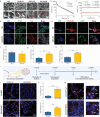
Schematic illustration of the inorganic-biomaterials-based neural platform and the characterization of the biological effects of LCS microspheres. (a) Schematic illustration of the preparation and application of an inorganic-biomaterials-based neural platform. Inorganic bioinks were composed of gelatin, gelatin methacryloyl (GelMA), Li-Ca-Si (LCS) microspheres and neural stem cells (NSCs). The 3D bioprinted neural constructs could serve as a versatile platform for promoting the regeneration of multiple tissues with functional recovery, including spinal cord injury repair, innervated bone regeneration and innervated muscle regeneration. (b and c) SEM images of LCS microspheres. (d) Size distribution of LCS microspheres. (e) X-ray diffraction (XRD) pattern of LCS microspheres. (f) Representative immunofluorescence staining images of Nestin proteins expression in NSCs after 5 days of culture. (g) Quantitative analysis of the mean fluorescence intensity of Nestin in Blank and LCS-250 groups (n = 5). (h) Representative immunofluorescence staining images of GFAP and Tuj1 proteins expression in NSCs after 5 days of culture. (i) Percentage of GFAP- and Tuj1-positive cells per field (n = 5). (j) Representative immunofluorescence staining images of MAP2 proteins after 10 days of culture. (k) Percentage of MAP2-positive cells per field (n = 5). (l) Schematic depiction of the stimulatory effects of LCS microspheres on the neural differentiation of NSCs. LCS bioceramic microspheres significantly stimulated the neuronal differentiation of NSCs and neuron maturation. Data are presented as the mean value ± SD. *P < 0.05, **P < 0.01, ***P < 0.001.
Figure 2.
Figure 2.
Characterization of the neuronal differentiation activity of 3D bioprinted neural constructs in vitro and in vivo. (a) SEM images of GG, GG-2LCS, GG-5LCS and GG-10LCS hydrogel inks. Red arrows indicate the incorporated LCS microspheres. (b) The shear-thinning properties of the hydrogel inks added with different contents of LCS. (c) The storage modulus (G′) and loss modulus (G′′) of these hydrogel inks with a frequency range of 0.1 to 10 Hz. (d) Representative double-immunofluorescence staining images of GFAP and Tuj1 of NSCs within GG and GG-5LCS constructs after 10 days of culture. (e) Representative immunofluorescence staining images of mature neuron marker MAP2 after 14 days of culture. (f–h) The percentage (normalized to nuclei) of GFAP- (f), Tuj1- (g) and MAP2- (h) positive cells per field in GG and GG-5LCS groups after (n = 4). (i) Schematic diagram of the procedure of subcutaneous implantation. (j) Representative immunofluorescence staining images of the neuronal markers Tuj1 and MAP2 in the constructs, 14 days post-implantation. (k and l) The quantitative analysis of Tuj1- and MAP2-positive areas per field in vivo (n = 4). (m) Double-immunofluorescence staining of GFP/Tuj1 and GFP/MAP2 demonstrated the survival, neuronal differentiation and maturation of exogenous NSCs within the constructs after being implanted for 14 days. White arrows indicate the co-localization. NSCs within the 3D bioprinted neural constructs exhibited superior survival, neuronal differentiation and neuron maturation activities under the stimulation of LCS microspheres both in vitro and in vivo. Data are presented as the mean value ± SD. *P < 0.05, **P < 0.01, ***P < 0.001.
Figure 3.
Figure 3.
RNA-seq analysis of the mechanism of LCS microspheres for the neuronal differentiation of NSCs. (a) A volcano plot of differentially expressed genes (DEGs) of NSCs within the bioprinted GG-LCS-NSC and GG-NSC constructs, which were identified by RNA-sequencing with the cutoff: p-value < 0.05 and |log2 Fold Change| > 1. The 1338 down-regulated genes were marked in blue and the 185 up-regulated genes were marked in red. (b) Heat map plot of the top 30 differentially up-regulated and down-regulated genes of GG-LCS-NSC and GG-NSC groups. (c) Gene ontology (GO) enrichment of the biological function of DEGs. (d) Kyoto Encyclopedia of Genes and Genomes (KEGG) analysis of the signal pathway enriched by the DEGs. (e) Fold changes of the DEGs enriched in PI3K-AKT signal pathway. (f) RT-qPCR analysis results of the DEGs, which were involved in the PI3K-AKT signal pathway (n = 3). (g) Western blot analysis of the expression of proteins related to the PI3K-Akt signaling pathway and neuronal differentiation, including p-PI3K, PI3K, p-AKT, AKT, downstream target proteins GSK-3β and neuron marker β-III tubulin. (h) A schematic illustration of the potential underlying mechanism of LCS microspheres for the neuronal differentiation of NSCs within bioprinted constructs. LCS microspheres promote the neuronal differentiation of NSCs and neuron maturation within the bioprinted constructs via activating the PI3K-AKT signal pathway. Data are presented as the mean value ± SD. *P < 0.05, **P < 0.01, ***P < 0.001.
Figure 4.
Figure 4.
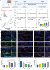
Therapeutic effects of 3D bioprinted neural constructs on the repair and functional recovery of spinal cord injury (SCI). (a) Schematic diagram of the procedure for SCI repair experiments. (b) The BBB score of SCI rats in each group. (c) Electrophysiological analysis of SCI rats in different groups after 8 weeks of post-surgery. (d) The average amplitudes of MEP of all groups after 8 weeks of surgery (n = 3). (e and f) Representative immunofluorescence staining images of Tuj1 (e) and GFAP (f) of the longitudinal sections of spinal cords, for evaluating neuronal regeneration. (g) Representative immunofluorescence staining images of CD31 of the longitudinal sections of spinal cords, for evaluating angiogenesis. (h–j) Statistical analysis of the Tuj1- (h), GFAP- (i) and CD31- (j) positive areas in the lesion regions (n = 5). 3D bioprinted GG-LCS-NSC neural constructs promoted the repairment and motor functional recovery of the spinal cord in a complete transection SCI model. Data are presented as the mean value ± SD. *P < 0.05, **P < 0.01, ***P < 0.001.
Figure 5.
Figure 5.
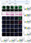
3D bioprinted neural constructs promoting bone regeneration in a cranial defect model. (a) A schematic diagram of the procedure for bone regeneration experiments. (b) Representative micro-CT reconstruction images of the bone defect regions in both the coronal and sagittal view. (c–f) Statistical analysis of the BV/TV (c), BMD (d), Tb.N (e) and Tb.Sp (f) values of regenerated bones (n = 6). (g) Microscope images of the H&E staining of the regenerated bone tissues at 8 weeks of implantation. (h and i) Representative images of osteogenic marker OCN (h) and OPN (i) for evaluating bone formation. (j and k) Representative images of neural marker NF (j) and CGRP (k) for evaluating innervation. (l) Representative images of angiogenic marker CD31 for evaluating vascularization. (m–q) Statistical analysis of the OCN (m), OPN (n), NF (o), CGRP (p) and CD31 (q) positive areas in the bone defect regions (n = 5). 3D bioprinted GG-LCS-NSC neural constructs significantly promoted bone regeneration with innervation and vascularization under the synergistic effects of NSCs and LCS bioceramic. Data are presented as the mean value ± SD. *P < 0.05, **P < 0.01, ***P < 0.001.
Figure 6.
Figure 6.
3D bioprinted neural constructs promoting muscle regeneration in a rat tibialis anterior (TA) muscle defect model. (a) A schematic diagram of the procedure for skeletal muscle regeneration experiments. (b) Representative optical images of the retrieved TA muscles after 8 weeks of implantation. (c) The weight of retrieved TA muscles (% of contralateral) after 8 weeks of implantation (n = 6). (d) Representative images of H&E staining and Masson's trichrome staining of the regenerated TA muscles. (e) Representative images of myogenic marker MHC, for evaluating the regeneration and maturation of newly formed myofibers. (f) Representative images of angiogenic marker CD31 for assessing vascularization. (g) Representative images of the double-immunofluorescence staining of AchRs and NF for evaluating neural integration. (h–k) Statistical analysis of the MHC (h), CD31 (i), NF (j) and AchRs (k) positive areas in the TA muscle defect regions (n = 5). 3D bioprinted GG-LCS-NSC neural constructs promoted myofiber formation and maturation, vascularization, and neural integration of the regenerated muscles. Data are presented as the mean value ± SD. *P < 0.05, **P < 0.01, ***P < 0.001.

Similar articles

Cited by

References

    1. Kerkman JN, Daffertshofer A, Gollo LL et al. Network structure of the human musculoskeletal system shapes neural interactions on multiple time scales. Sci Adv 2018; 4: eaat0497.10.1126/sciadv.aat0497 - DOI - PMC - PubMed
    1. Elefteriou F. Impact of the autonomic nervous system on the skeleton. Physiol Rev 2018; 98: 1083–112.10.1152/physrev.00014.2017 - DOI - PMC - PubMed
    1. Varadarajan SG, Hunyara JL, Hamilton NR et al. Central nervous system regeneration. Cell 2022; 185: 77–94.10.1016/j.cell.2021.10.029 - DOI - PMC - PubMed
    1. Zhang H, Wu C. 3D printing of biomaterials for vascularized and innervated tissue regeneration. Inter J Bioprint 2023; 9: 1–21.10.18063/ijb.706 - DOI - PMC - PubMed
    1. Tomlinson RE, Li Z, Zhang Q et al. NGF-TrkA signaling by sensory nerves coordinates the vascularization and ossification of developing endochondral bone. Cell Rep 2016; 16: 2723–35.10.1016/j.celrep.2016.08.002 - DOI - PMC - PubMed