[Preprint]. 2024 Sep 23:2024.02.28.582593.
doi: 10.1101/2024.02.28.582593.

Electrophysiological dynamics of salience, default mode, and frontoparietal networks during episodic memory formation and recall: A multi-experiment iEEG replication

Affiliations

Electrophysiological dynamics of salience, default mode, and frontoparietal networks during episodic memory formation and recall: A multi-experiment iEEG replication

Anup Das et al. bioRxiv. .

Update in

Abstract

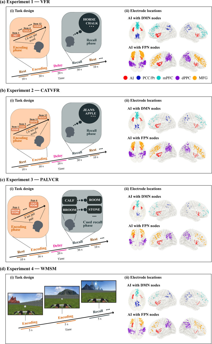
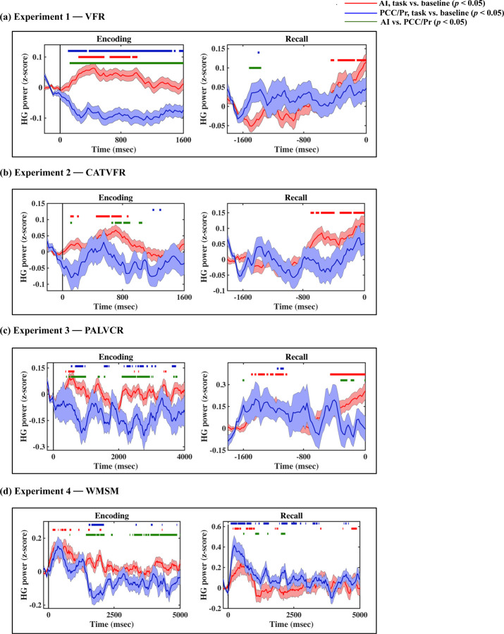
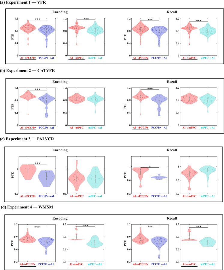
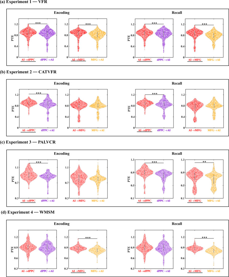
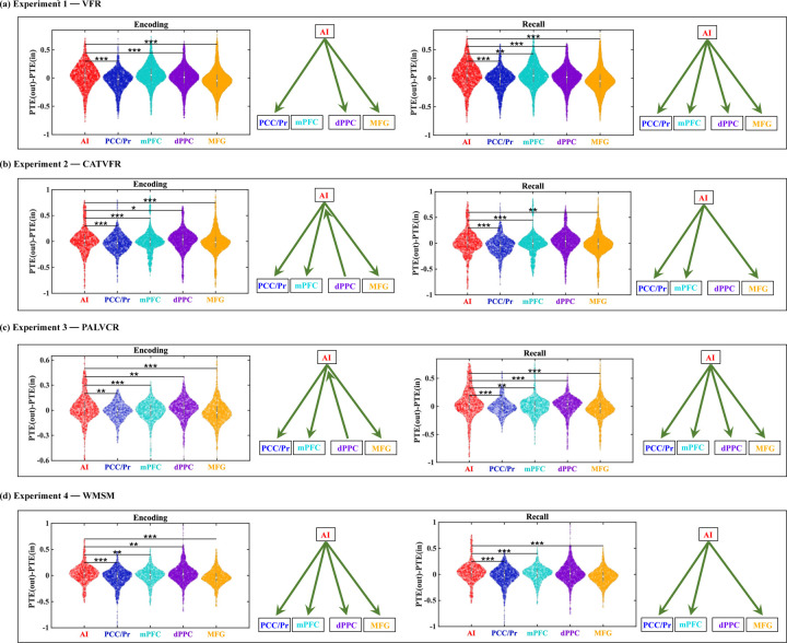
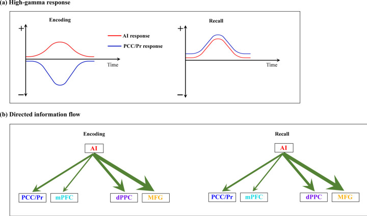
Dynamic interactions between large-scale brain networks underpin human cognitive processes, but their electrophysiological mechanisms remain elusive. The triple network model, encompassing the salience (SN), default mode (DMN), and frontoparietal (FPN) networks, provides a framework for understanding these interactions. We analyzed intracranial EEG recordings from 177 participants across four diverse episodic memory experiments, each involving encoding as well as recall phases. Phase transfer entropy analysis revealed consistently higher directed information flow from the anterior insula (AI), a key SN node, to both DMN and FPN nodes. This directed influence was significantly stronger during memory tasks compared to resting-state, highlighting the AI's task-specific role in coordinating large-scale network interactions. This pattern persisted across externally-driven memory encoding and internally-governed free recall. Control analyses using the inferior frontal gyrus (IFG) showed an inverse pattern, with DMN and FPN exerting higher influence on IFG, underscoring the AI's unique role. We observed task-specific suppression of high-gamma power in the posterior cingulate cortex/precuneus node of the DMN during memory encoding, but not recall. Crucially, these results were replicated across all four experiments spanning verbal and spatial memory domains with high Bayes replication factors. Our findings advance understanding of how coordinated neural network interactions support memory processes, highlighting the AI's critical role in orchestrating large-scale brain network dynamics during both memory encoding and retrieval. By elucidating the electrophysiological basis of triple network interactions in episodic memory, our study provides insights into neural circuit dynamics underlying memory function and offer a framework for investigating network disruptions in memory-related disorders.

Keywords: Human intracranial EEG; attentional control; default-mode network; episodic memory; frontoparietal network; human insula; salience network; triple-network model.


Conflict of interest statement

Conflict of interest statement: The authors declare no competing financial interests.

Figures

Figure 1.
Figure 1.. Task design of the encoding and recall periods of the memory experiments, and iEEG recording sites in AI, with DMN and FPN nodes.
(a) Experiment 1, Verbal free recall (VFR): (i) Task design of memory encoding and recall periods of the verbal free recall experiment (see Methods for details). Participants were first presented with a list of words in the encoding block and asked to recall as many as possible from the original list after a short delay. (ii) Electrode locations for AI with DMN nodes (top panel) and AI with FPN nodes (bottom panel), in the verbal free recall experiment. Proportion of electrodes for AI, PCC/Pr, mPFC, dPPC, and MFG were 9%, 8%, 19%, 32%, and 32% respectively, in the VFR experiment. (b) Experiment 2, Categorized verbal free recall (CATVFR): (i) Task design of memory encoding and recall periods of the categorized verbal free recall experiment (see Methods for details). Participants were presented with a list of words with consecutive pairs of words from a specific category (for example, JEANS-COAT, GRAPE-PEACH, etc.) in the encoding block and subsequently asked to recall as many as possible from the original list after a short delay. (ii) Electrode locations for AI with DMN nodes (top panel) and AI with FPN nodes (bottom panel), in the categorized verbal free recall experiment. Proportion of electrodes for AI, PCC/Pr, mPFC, dPPC, and MFG were 10%, 7%, 11%, 35%, and 37% respectively, in the CATVFR experiment. (c) Experiment 3, Paired associates learning verbal cued recall (PALVCR): (i) Task design of memory encoding and recall periods of the paired associates learning verbal cued recall experiment (see Methods for details). Participants were first presented with a list of 6 word-pairs in the encoding block and after a short post-encoding delay, participants were shown a specific word-cue and asked to verbally recall the cued word from memory. (ii) Electrode locations for AI with DMN nodes (top panel) and AI with FPN nodes (bottom panel), in the paired associates learning verbal cued recall experiment. Proportion of electrodes for AI, PCC/Pr, mPFC, dPPC, and MFG were 14%, 5%, 13%, 33%, and 35% respectively, in the PALVCR experiment. (d) Experiment 4, Water maze spatial memory (WMSM): (i) Task design of memory encoding and recall periods of the water maze spatial memory experiment (see Methods for details). Participants were shown objects in various locations during the encoding period and asked to retrieve the location of the objects during the recall period. Modified from Jacobs et. al. (2018) with permission. (ii) Electrode locations for AI with DMN nodes (top panel) and AI with FPN nodes (bottom panel), in the water maze spatial memory experiment. Proportion of electrodes for AI, PCC/Pr, mPFC, dPPC, and MFG were 10%, 15%, 13%, 38%, and 24% respectively, in the WMSM experiment. Overall, proportion of electrodes for VFR, CATVFR, PALVCR, and WMSM experiments were 43%, 27%, 15%, and 15% respectively. AI: anterior insula, PCC: posterior cingulate cortex, Pr: precuneus, mPFC: medial prefrontal cortex, dPPC: dorsal posterior parietal cortex, MFG: middle frontal gyrus.
Figure 2.
Figure 2.. Anterior insula electrode locations (red) visualized on insular regions based on the atlas by Faillenot and colleagues (Faillenot, Heckemann, Frot, & Hammers, 2017).
Anterior insula is shown in blue, and posterior insula mask is shown in green (see Methods for details). This atlas is based on probabilistic analysis of the anatomy of the insula with demarcations of the AI based on three short dorsal gyri and the PI which encompasses two long and ventral gyri.
Figure 3.
Figure 3.. iEEG evoked response, quantified using high-gamma (HG) power, for AI (red) and PCC/precuneus (blue) during (a) VFR, (b) CATVFR, (c) PALVCR, and (d) WMSM experiments.
Green horizontal lines denote greater high-gamma power for AI compared to PCC/precuneus (ps < 0.05). Red horizontal lines denote increase of AI response compared to the resting baseline during the encoding and recall periods (ps < 0.05). Blue horizontal lines denote decrease of PCC/precuneus response compared to the baseline during the encoding periods and increase of PCC/precuneus response compared to the baseline during the recall periods (ps < 0.05).
Figure 4.
Figure 4.. Directed information flow between the anterior insula and the PCC/precuneus and mPFC nodes of the default mode network (DMN), across verbal and spatial memory domains, measured using phase transfer entropy (PTE).
(a) Experiment 1, VFR: The anterior insula showed higher directed information flow to the PCC/precuneus (AI ➜ PCC/Pr) compared to the reverse direction (PCC/Pr ➜ AI) (n=142) during both encoding and recall. The anterior insula also showed higher directed information flow to the mPFC (AI ➜ mPFC) compared to the reverse direction (mPFC ➜ AI) (n=112) during both memory encoding and recall. (b) Experiment 2, CATVFR: The anterior insula showed higher directed information flow to the PCC/precuneus (AI ➜ PCC/Pr) compared to the reverse direction (PCC/Pr ➜ AI) (n=46) during both encoding and recall. (c) Experiment 3, PALVCR: The anterior insula showed higher directed information flow to the PCC/precuneus (AI ➜ PCC/Pr) compared to the reverse direction (PCC/Pr ➜ AI) (n=10) during both encoding and recall. (d) Experiment 4, WMSM: The anterior insula showed higher directed information flow to PCC/precuneus (AI ➜ PCC/Pr) than the reverse (PCC/Pr ➜ AI) (n=91), during both spatial memory encoding and recall. The anterior insula also showed higher directed information flow to mPFC (AI ➜ mPFC) than the reverse (mPFC ➜ AI) (n=23), during both spatial memory encoding and recall. In each panel, the direction for which PTE is higher, is underlined. White dot in each violin plot represents median PTE across electrode pairs. *** p < 0.001, * p < 0.05.
Figure 5.
Figure 5.. Directed information flow between the anterior insula and the dPPC and MFG nodes of the frontoparietal network (FPN), across verbal and spatial memory domains.
(a) Experiment 1, VFR: The anterior insula showed higher directed information flow to the dorsal PPC (AI ➜ dPPC) compared to the reverse direction (dPPC ➜ AI) (n=586) during both encoding and recall. The anterior insula also showed higher directed information flow to the MFG (AI ➜ MFG) compared to the reverse direction (MFG ➜ AI) (n=642) during both memory encoding and recall. (b) Experiment 2, CATVFR: The anterior insula showed higher directed information flow to the dorsal PPC (AI ➜ dPPC) compared to the reverse direction (dPPC ➜ AI) (n=327) during both encoding and recall. (c) Experiment 3, PALVCR: The anterior insula showed higher directed information flow to the dorsal PPC (AI ➜ dPPC) compared to the reverse direction (dPPC ➜ AI) (n=242) during both encoding and recall. The anterior insula also showed higher directed information flow to the MFG (AI ➜ MFG) compared to the reverse direction (MFG ➜ AI) (n=362) during memory recall. (d) Experiment 4, WMSM: The anterior insula showed higher directed information flow to MFG (AI ➜ MFG) than the reverse (MFG ➜ AI) (n=177), during both spatial memory encoding and recall. In each panel, the direction for which PTE is higher, is underlined. *** p < 0.001, ** p < 0.01.
Figure 6.
Figure 6.. The anterior insula is an outflow hub in its interactions with the DMN and FPN, during encoding and recall periods, and across memory experiments.
In each panel, the net direction of information flow between the AI and the DMN and FPN nodes are indicated by green arrows on the right. *** p < 0.001, ** p < 0.01, * p < 0.05.
Figure 7.
Figure 7.. Schematic illustration of key findings related to the intracranial electrophysiology of the triple network model in human episodic memory.
(a) High-gamma response: Our analysis of local neuronal activity revealed consistent suppression of high-gamma power in the PCC/precuneus compared to the AI during encoding periods across all four episodic memory experiments. We did not consistently observe any significant differences in high-gamma band power between AI and the mPFC node of the DMN or the dPPC and MFG nodes of the FPN during the encoding periods across the four episodic memory experiments. In contrast, we detected similar high-gamma band power in the PCC/precuneus relative to the AI during the recall periods. (b) Directed information flow: Despite variable patterns of local activation and suppression across DMN and FPN nodes during memory encoding and recall, we found stronger directed influence (denoted by green arrows, thickness of arrows denotes degree of replicability across experiments, see Table 1) by the AI on both the DMN as well as the FPN nodes than the reverse, across all four memory experiments, and during both encoding and recall periods.

Similar articles

References

    1. Andermane N., Joensen B. H., & Horner A. J. (2021). Forgetting across a hierarchy of episodic representations. Curr Opin Neurobiol, 67, 50–57. doi:10.1016/j.conb.2020.08.004 - DOI - PubMed
    1. Andrews-Hanna J. R. (2012). The brain’s default network and its adaptive role in internal mentation. Neuroscientist, 18(3), 251–270. doi:10.1177/1073858411403316 - DOI - PMC - PubMed
    1. Atkinson R. C., & Shiffrin R. M. (1968). Human memory: a proposed system and its control processes. Psychology of Learning and Motivation, 2, 89–195.
    1. Badre D., Poldrack R. A., Paré-Blagoev E. J., Insler R. Z., & Wagner A. D. (2005). Dissociable controlled retrieval and generalized selection mechanisms in ventrolateral prefrontal cortex. Neuron, 47(6), 907–918. doi:10.1016/j.neuron.2005.07.023 - DOI - PubMed
    1. Badre D., & Wagner A. D. (2007). Left ventrolateral prefrontal cortex and the cognitive control of memory. Neuropsychologia, 45(13), 2883–2901. doi:10.1016/j.neuropsychologia.2007.06.015 - DOI - PubMed

Publication types