[Preprint]. 2024 Feb 21:rs.3.rs-3941185.
doi: 10.21203/rs.3.rs-3941185/v1.

Integrative structural analysis of Pseudomonas phage DEV reveals a genome ejection motor

Affiliations

Integrative structural analysis of Pseudomonas phage DEV reveals a genome ejection motor

Gino Cingolani et al. Res Sq. .

Update in

Abstract

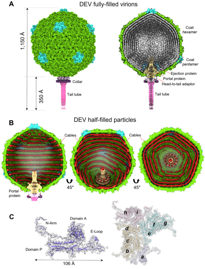
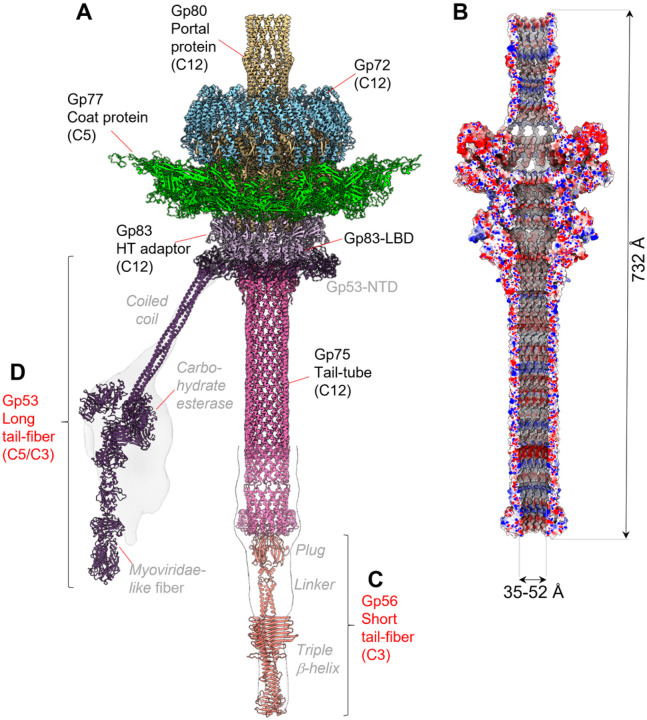
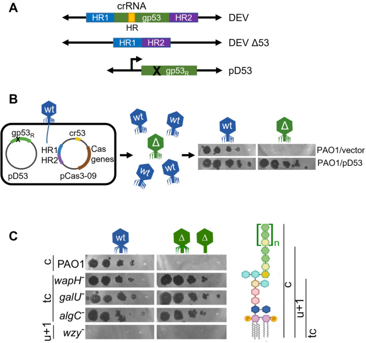
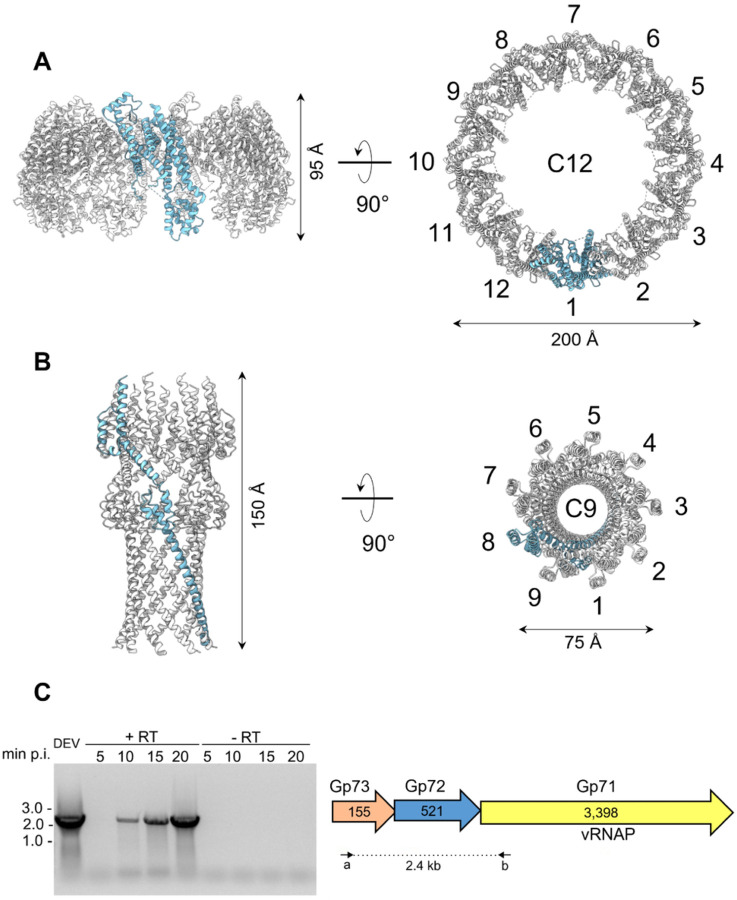
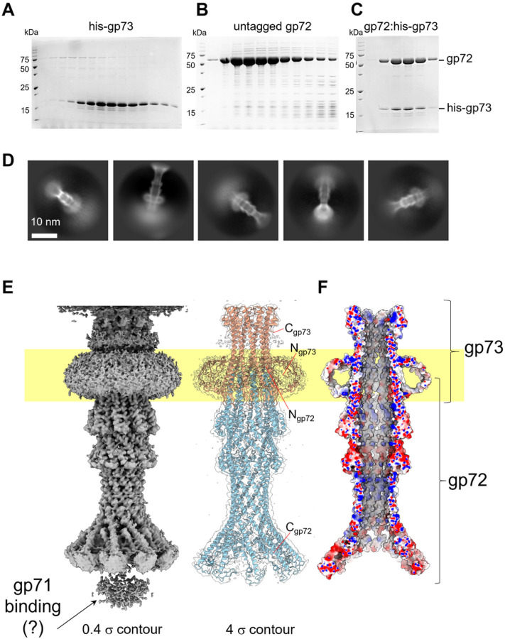
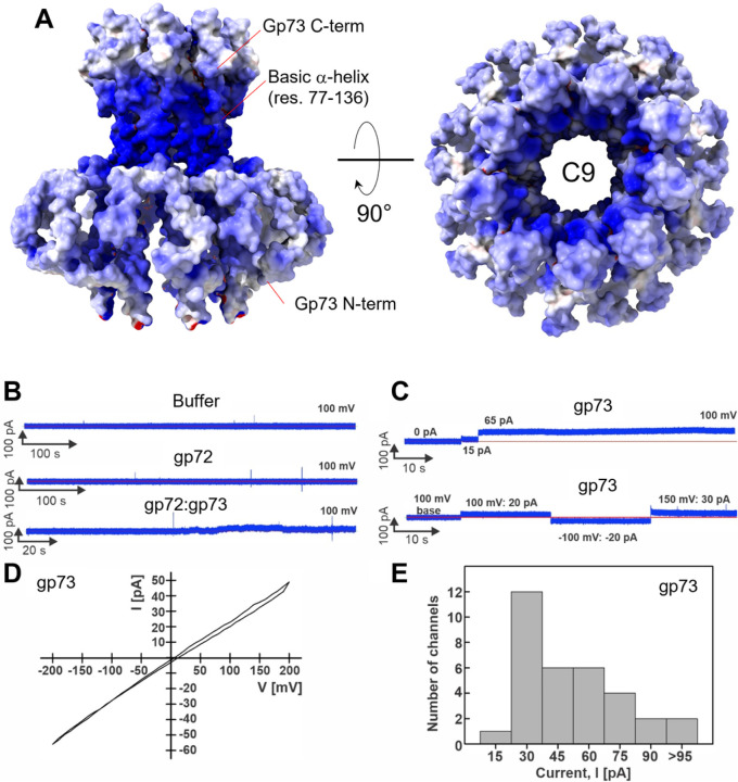
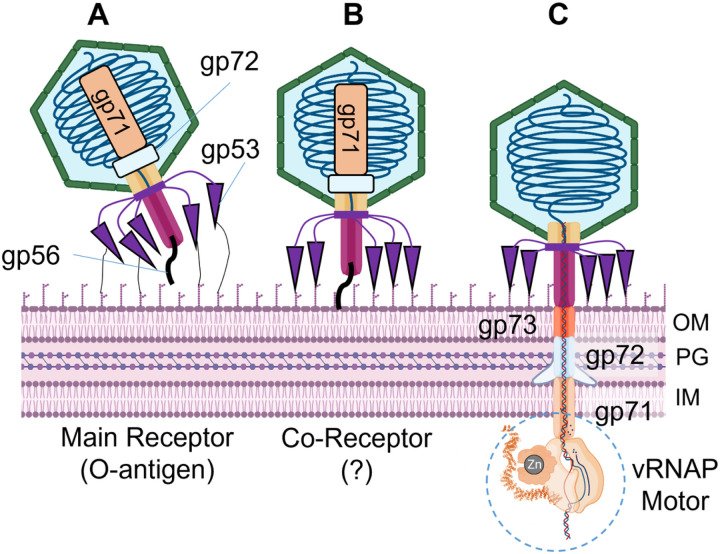
DEV is an obligatory lytic Pseudomonas phage of the N4-like genus, recently reclassified as Schitoviridae. The DEV genome encodes 91 ORFs, including a 3,398 amino acid virion-associated RNA polymerase. Here, we describe the complete architecture of DEV, determined using a combination of cryo-electron microscopy localized reconstruction, biochemical methods, and genetic knockouts. We built de novo structures of all capsid factors and tail components involved in host attachment. We demonstrate that DEV long tail fibers are essential for infection of Pseudomonas aeruginosa and dispensable for infecting mutants with a truncated lipopolysaccharide devoid of the O-antigen. We identified DEV ejection proteins and, unexpectedly, found that the giant DEV RNA polymerase, the hallmark of the Schitoviridae family, is an ejection protein. We propose that DEV ejection proteins form a genome ejection motor across the host cell envelope and that these structural principles are conserved in all Schitoviridae.


Conflict of interest statement

COMPETING INTERESTS STATEMENT The authors declare no competing interests.

Figures

Figure 1
Figure 1. Cryo-EM analysis of the Pseudomonas phage DEV.
(A) Asymmetric cryo-EM reconstruction of DEV FF virion in a side (left) and cutout (right) view. The T = 9 icosahedral shell is colored light green (hexamers) and cyan (pentons). (B) Asymmetric cryo-EM reconstructions of DEV HF particle. From left to right, three cutout views of the capsid are shown rotated in 45 Å increments. The cable density assigned to dsDNA is colored red. (C) (Left) DEV coat protein gp77 tertiary structure overlaid to a 3.3 Å C5-averaged localized reconstruction of the mature head map contoured at 5sabove background. (Right) Overview of DEV T = 9 icosahedral asymmetric unit comprising nine coat proteins labeled (a−i).
Figure 2
Figure 2. DEV tail apparatus.
(A). Composite ribbon diagram of DEV tail reconstructed from FF virions. Tail factors identified de novo in the C12 localized reconstruction include the portal protein gp80 (yellow), the ejection protein gp72 (blue), the HT-adaptor gp83 (light purple), and the tail tube gp75 (magenta). (B) Cross section of an electrostatic surface representation of the DEV tail channel. Red, blue, and white represent negative, positive, and neutral charges near the surface. (C–D) AlphaFold models for the short-tail fiber gp56 and long-tail fiber gp53 overlaid to low-resolution localized reconstructions shown as semitransparent surfaces. Individual tail factors are color-coded, as in panel A.
Figure 3
Figure 3. Topology and composition of DEV collar.
(A) A low-resolution C5 map of the DEV mature virion visualized from the bottom of the tail apparatus shows density for five appendages protruding from the collar and assigned to the tail fibers gp53. (B) A C15 symmetrized density of the DEV collar is visualized at a high contour (5s). The AlphaFold model of the tail fiber gp53-NTB was docked into the density. Fifteen copies of gp53-NTB fill the collar density with no clashes.
Figure 4
Figure 4. Role of DEV long fiber gp53 in host attachment.
(A) Structure of the gp53 locus in DEV and DEV Δ53 phages and the pD53 plasmid. crRNA HR, region targeted by cr-RNA53 expressed by pCas3-09; HR1 and HR2, homology regions cloned in pCas3-09 plasmid; gp53R, cr-RNA53 resistant gp53 allele cloned in pD53. (B) Outline of DEV mutagenesis. Infection with DEV of PAO1 carrying pCas3-09 and pD53 produces a genetically mixed phage progeny with wt (in blue) and Δ53 (in green) virions. Unlike gp53+ DEV, Δgp53 mutants grow on PAO1 carrying pD53 (pD53) but not on PAO1 containing the empty vector pGM931 (EV). (C) DEV Δgp53 growth on mutants with LPS defects. Serial dilutions (x 10) of DEV or Δgp53 were replicated on PAO1 and the indicated PAO1 mutants with defective LPS. On the right, the structure of PAO1 LPS (c, capped) and the LPS portions present in the mutant LPS variants. tc, truncated core; u+1, uncapped LPS + one O-antigen repeat.
Figure 5
Figure 5. Quaternary structures of DEV ejection protein gp72 pre- and post-ejection.
(A) The quaternary structure of DEV gp72 from FF virions determined in situ. Twelve gp72 subunits surround the portal protein, generating a ~200 Å wide ring. (B) Cryo-EM structure of the recombinant nonameric gp72 determined at 3.65 Å resolution in the post-ejection conformation. In panels A and B, only one protomer is colored in cyan, whereas all other subunits are light gray. (C) DEV gp71, gp72, and gp73 genes are co-transcribed as an operon. (Left panel) Agarose gel electrophoresis of RT-PCR products. RNA samples extracted from PAO1 cultures at different time points post-infection (p.i.) with DEV (e.g., 5, 10, 15, 20 minutes) were reverse-transcribed (+RT) or not (negative control, −RT) and used as templates for PCR amplification. Migration of MW (kb) markers is shown on the left. (Right panel) Schematic diagram of DEV ORFs encoding gp71, gp72, and gp73. Arrows represent the position of oligonucleotides used for amplification, yielding a 2.4 kb long amplification product.
Figure 6
Figure 6. DEV ejection proteins gp72 and gp73 form a tube-shaped complex.
SDS-PAGE analysis of purified (A) hisgp73 solubilized from membranes; (B) gp72 expressed under native conditions; (C) gel filtration fractions containing the gp72:gp73 complex. (D) Representative 2D class averages of the gp72:gp73 complex. (E) 3D reconstruction of the gp72:gp73 complex visualized at low (left) and high (right) contours. The atomic models of gp73 and gp72 are overlaid to the semitransparent density calculated at 3.15 Å resolution. In yellow is the putative position of the bacterium’s outer membrane. (F) The cross-section of an electrostatic surface representation of gp72:gp73 shows the lumen and surface charge inside the channel. Red, blue, and white represent negative, positive, and neutral charges near the surface.
Figure 7
Figure 7. Lipid bilayer experiments with purified DEV ejection protein gp73.
(A) The electrostatic surface representation of nonameric gp73 reveals a significant positive charge, mainly in the a-helical core. (B–C) Lipid bilayer experiments were performed at 100 mV applied potential in diphytanoylphosphotidylcholine (DPhPC) membranes bathed in 1 M KCl, 10 mM HEPES, pH 7.4 electrolyte. The protein samples were added to the grounded trans side of the cuvette, which had 100 μm SU-8 aperture. (B) Representative current traces. Top: 15 μl protein buffer in the cuvette. Six membranes were recorded with 1 – 15 μl of the protein buffer, and no activity of the buffer was observed. Middle: gp72 current trace. Seven membranes with up to 24 μg of gp72 in the cuvette were recorded, and no channel activity was observed. Bottom: gp72:gp73 complex. 19 membranes were recorded, and only one shown here had 10 – 20 pA fluctuations around the baseline when 10 μg of protein sample was in the cuvette. (C) Representative current traces of gp73. Top: Two insertions of gp73 (750 ng protein in the cuvette) with amplitudes of 15 pA, and 65 pA. Bottom: Continuous current trace of a single gp73 insertion (900 ng in the cuvette) at indicated voltages. (D) Current-voltage curve of one gp73 pore inserted in the DPhPC membrane at a voltage range of −200 to 200 mV. (E) Histogram of single-channel current amplitudes of gp73 at 100 mV. A total of 33 channels were observed with a mode current of 30 pA.
Figure 8
Figure 8. Model for DEV absorption onto P. aeruginosa surface and genome ejection.
Three proposed steps of infection are shown: each step is accompanied by distinct conformations of the long and short-tail fibers. (A) DEV interacts with the host O-antigen through flexible long-tail fibers (gp53), possibly reorienting the virion to land perpendicular to the OM. (B) The short-tail fiber gp56 interacts with a secondary receptor in the bacterium OM, triggering a conformational change that releases the short fiber. (C) The ejection proteins gp73, gp72, and gp71 are expelled into the bacterium cell envelope where gp72 forms an OM pore, gp73 spans the periplasm, and gp71 crosses the IM, projecting a large vRNAP motor into the bacterial cytoplasm, that begins pulling the viral genome inside the host. PG = peptidoglycan

Similar articles

References

    1. Wittmann J. et al. From Orphan Phage to a Proposed New Family-the Diversity of N4-Like Viruses. Antibiotics (Basel) 9(2020). - PMC - PubMed
    1. Menon N.D. et al. A Novel N4-Like Bacteriophage Isolated from a Wastewater Source in South India with Activity against Several Multidrug-Resistant Clinical Pseudomonas aeruginosa Isolates. mSphere 6(2021). - PMC - PubMed
    1. Shi X. et al. Characterization and Complete Genome Analysis of Pseudomonas aeruginosa Bacteriophage vB_PaeP_LP14 Belonging to Genus Litunavirus. Curr Microbiol 77, 2465–2474 (2020). - PubMed
    1. Lerdsittikul V. et al. A novel virulent Litunavirus phage possesses therapeutic value against multidrug resistant Pseudomonas aeruginosa. Sci Rep 12, 21193 (2022). - PMC - PubMed
    1. Tajuddin S. et al. Genomic analysis and biological characterization of a novel Schitoviridae phage infecting Vibrio alginolyticus. Appl Microbiol Biotechnol 107, 749–768 (2023). - PubMed

Publication types