[Preprint]. 2024 Feb 19:2024.02.17.580812.
doi: 10.1101/2024.02.17.580812.

Inhibition of Soluble Epoxide Hydrolase Reduces Inflammation and Myocardial Injury in Arrhythmogenic Cardiomyopathy

Affiliations

Inhibition of Soluble Epoxide Hydrolase Reduces Inflammation and Myocardial Injury in Arrhythmogenic Cardiomyopathy

Dipak Panigrahy et al. bioRxiv. .

Update in

Abstract

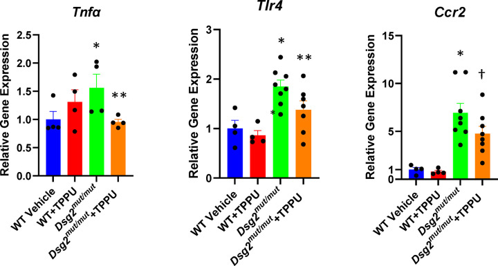
Previous studies have implicated persistent innate immune signaling in the pathogenesis of arrhythmogenic cardiomyopathy (ACM), a familial non-ischemic heart muscle disease characterized by life-threatening arrhythmias and progressive myocardial injury. Here, we provide new evidence implicating inflammatory lipid autocoids in ACM. We show that specialized pro-resolving lipid mediators are reduced in hearts of Dsg2mut/mut mice, a well characterized mouse model of ACM. We also found that ACM disease features can be reversed in rat ventricular myocytes expressing mutant JUP by the pro-resolving epoxy fatty acid (EpFA) 14,15-eicosatrienoic acid (14-15-EET), whereas 14,15-EE-5(Z)E which antagonizes actions of the putative 14,15-EET receptor, intensified nuclear accumulation of the desmosomal protein plakoglobin. Soluble epoxide hydrolase (sEH), an enzyme that rapidly converts pro-resolving EpFAs into polar, far less active or even pro-inflammatory diols, is highly expressed in cardiac myocytes in Dsg2mut/mut mice. Inhibition of sEH prevented progression of myocardial injury in Dsg2mut/mut mice and led to recovery of contractile function. This was associated with reduced myocardial expression of genes involved in the innate immune response and fewer pro-inflammatory macrophages expressing CCR2, which mediate myocardial injury in Dsg2mut/mut mice. These results suggest that pro-inflammatory eicosanoids contribute to the pathogenesis of ACM and, further, that inhibition of sEH may be an effective, mechanism-based therapy for ACM patients.


Conflict of interest statement

Author Conflicts of Interest Disclosures: Dr. Saffitz is a consultant to Implicit Biosciences and Rejuvenate Bio. Dr. Hammock holds patents related to the commercial development of soluble epoxide hydrolase inhibitors for cardiovascular disease. He is Chief Scientific Officer of EicOsis Human Health, currently in human 1b safety trials of the soluble epoxide hydrolase inhibitor EC5026. Dr. Lavine is a consultant for Kiniksa, Cytokinetics, Implicit Biosciences, and SUN Pharmaceuticals. Other authors have no relevant conflicts or financial relationships to disclose.

Figures

Figure 1:
Figure 1:
A. Reduced levels of specialized pro-resolving mediators resolvin D1, maresin 1 and resolvin E2 in hearts of 16-week-old Dsg2mut/mut compared to wildtype (WT) mice measured by ELISA, and of GPR18 in hearts of 16-week-old Dsg2mut/mut measured by western blotting. A sample of spleen was used as a positive control in the western blot; * p<0.05 vs. WT by Mann Whitney U test. B. qPCR showing increased expression of the endoplasmic reticulum chaperone gene BiP and the protein folding protein disulfide isomerase gene Pdi, both markers of endoplasmic reticulum stress. Gene expression values in wildtype samples were normalized to 1; values in Dsg2mut/mut samples are shown as relative levels; * p<0.05 vs. WT by Mann Whitney U test.
Figure 1:
Figure 1:
A. Reduced levels of specialized pro-resolving mediators resolvin D1, maresin 1 and resolvin E2 in hearts of 16-week-old Dsg2mut/mut compared to wildtype (WT) mice measured by ELISA, and of GPR18 in hearts of 16-week-old Dsg2mut/mut measured by western blotting. A sample of spleen was used as a positive control in the western blot; * p<0.05 vs. WT by Mann Whitney U test. B. qPCR showing increased expression of the endoplasmic reticulum chaperone gene BiP and the protein folding protein disulfide isomerase gene Pdi, both markers of endoplasmic reticulum stress. Gene expression values in wildtype samples were normalized to 1; values in Dsg2mut/mut samples are shown as relative levels; * p<0.05 vs. WT by Mann Whitney U test.
Figure 2.
Figure 2.
Effects of 14,15-eicosatrienoic acid (14,15-EET) and 14,15-eicosa-5(Z)-enoic acid (14,15-EEZE) on the distribution of immunoreactive signals for plakoglobin, connexin43 (Cx43) and RelA/p65 in primary cultures of wildtype (WT) neonatal rat ventricular myocytes and myocyes transfected to express the ACM disease allele JUP2157del2.
Figure 3.
Figure 3.
UMAPs showing populations of cells isolated from hearts of 16-week-old Dsg2mut/mut mice and the relative density of Ephx2 expression in each population.
Figure 4.
Figure 4.
Effects of 1 μM 1-trifluoro-methoxy-phenyl-3-(1-propionylpiperidin-4-yl) urea (TPPU) and 500 nM 4-(5-phenyl-3-{3-[3-(4-trifluoromethylphenyl)-ureido]-propyl}-pyrazol-1-yl)-benzenesulfonamide (PTUPB) on the distribution of of immunoreactive signals for plakoglobin, connexin43 (Cx43) and RelA/p65 in primary cultures of wildtype (WT) neonatal rat ventricular myocytes and myocyes transfected to express the ACM disease allele JUP2157del2.
Figure 5.
Figure 5.
Effects of 1-trifluoro-methoxy-phenyl-3-(1-propionylpiperidin-4-yl) urea (TPPU) on left ventricular ejection fraction (A) and fractional shortening (B) in wildtype (WT) and Dsg2mut/mut mice. Baseline echocardiography was performed in 9-week-old mice and then repeated after treatment for 4 weeks with TPPU or vehicle (Veh). Data are shown for each group (left) and each individual animal (right); * p<0.0001 vs. WT-Veh by multiple comparisons ANOVA.
Figure 5.
Figure 5.
Effects of 1-trifluoro-methoxy-phenyl-3-(1-propionylpiperidin-4-yl) urea (TPPU) on left ventricular ejection fraction (A) and fractional shortening (B) in wildtype (WT) and Dsg2mut/mut mice. Baseline echocardiography was performed in 9-week-old mice and then repeated after treatment for 4 weeks with TPPU or vehicle (Veh). Data are shown for each group (left) and each individual animal (right); * p<0.0001 vs. WT-Veh by multiple comparisons ANOVA.
Figure 6.
Figure 6.
Effects of 1-trifluoro-methoxy-phenyl-3-(1-propionylpiperidin-4-yl) urea (TPPU) on the amount of myocardial fibrosis (A) and the number of cells expressing CCR2 (B) in wildtype (WT) and Dsg2mut/mut mice (positive cells identified by arrows in representative immunostained tissue sections). Hearts were excised from animals after treatment for 4 weeks with TPPU or vehicle (Veh) and analyzed by histology in trichrome stained sections or by immunohistochemistry in sections stained with an anti-CCR2 antibody; * p<0.05 vs. WT; ** p<0.05 vs. vehicle-treated Dsg2mut/mut mice by multiple T test.
Figure 6.
Figure 6.
Effects of 1-trifluoro-methoxy-phenyl-3-(1-propionylpiperidin-4-yl) urea (TPPU) on the amount of myocardial fibrosis (A) and the number of cells expressing CCR2 (B) in wildtype (WT) and Dsg2mut/mut mice (positive cells identified by arrows in representative immunostained tissue sections). Hearts were excised from animals after treatment for 4 weeks with TPPU or vehicle (Veh) and analyzed by histology in trichrome stained sections or by immunohistochemistry in sections stained with an anti-CCR2 antibody; * p<0.05 vs. WT; ** p<0.05 vs. vehicle-treated Dsg2mut/mut mice by multiple T test.
Figure 7.
Figure 7.
Effects of 1-trifluoro-methoxy-phenyl-3-(1-propionylpiperidin-4-yl) urea (TPPU) on expression of Tnfα, Tlr4 and Ccr2 in hearts of wildtype (WT) and Dsg2mut/mut mice treated with TPPU or vehicle. Gene expression values in wildtype samples treated with vehicle were normalized to 1; values in all other groups are shown as relative levels; * p<0.05 vs. WT Veh; ** p<0.05 vs. Dsg2mut/mut mice; † p=0.0579 vs. Dsg2mut/mut mice, all by multiple comparisons ANOVA.

Similar articles

References

    1. Corrado D, Link M, Calkins H. Arrhythmogenic right ventricular cardiomyopathy. New Eng J Med, 2017; 376:61–72. - PubMed
    1. Corrado D, Basso C, Thiene G, McKenna WJ, Davies MJ, Fontaliran F, Nava A, Silvestri F, Blomstrom-Lundqvist C, Wlodarska EK, Fontaine G, Camerini F. Spectrum of clinico-pathologic manifestations of arrhythmogenic right ventricular cardiomyopathy/dysplasia: a multicenter study. J Am Coll Cardiol 1997; 30:1512–1520. - PubMed
    1. Chelko SP, Asimaki A, Lowenthal J, Bueno-Beti C, Bedja D, Scalco A, Amat-Codina N, Andersen P, Judge DP, Tung L, Saffitz JE. Therapeutic modulation of the immune response in arrhythmogenic cardiomyopathy. Circulation 2019; 140:1491–1505. - PMC - PubMed
    1. Chelko SP, Penna V, Engle M, Landim-Vieira ML, Cannon EN, Lavine K, Saffitz JE. Mechanisms of innate immune injury in arrhythmogenic cardiomyopathy. bioRxiv. 2023. Jul 13.2023.07.12.548682, doi: 10.1101/2023.07.12.548682. - DOI
    1. Hawthorne RN, Blazeski A, Lowenthal J, Kannan S, Teuben R, DiSilvestre D, MorrissetteMcAlmon J, Saffitz JE, Boheler KR, James CA, Chelko SP, Tomaselli G, Tung L. Altered electrical, biomolecular, and immunologic phenotypes in a novel patient-derived stem cell model of desmoglein-2 mutant ARVC. J Clin Med 2021; 10;10(14):3061. doi: 10.3390/jcm10143061. - DOI - PMC - PubMed

Publication types