[Preprint]. 2024 Mar 1:rs.3.rs-3915097.
doi: 10.21203/rs.3.rs-3915097/v1.

A tension offloading patch mitigates dermal fibrosis induced by pro-fibrotic skin injections

Affiliations

A tension offloading patch mitigates dermal fibrosis induced by pro-fibrotic skin injections

Heather E Talbott et al. Res Sq. .

Abstract

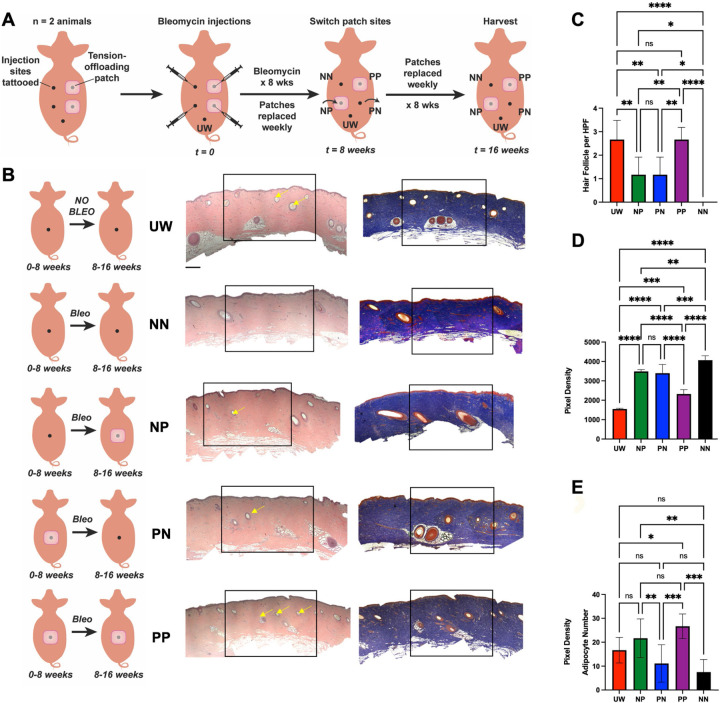
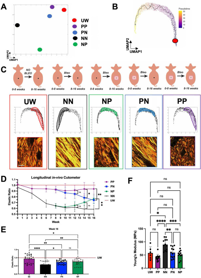
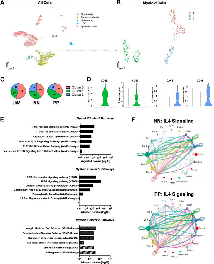
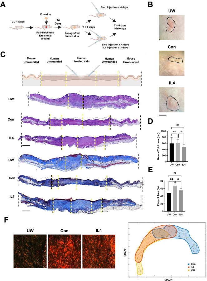
Skin fibrosis is a clinical problem with devastating impacts but limited treatment options. In the setting of diabetes, insulin administration often causes local dermal fibrosis, leading to a range of clinical sequelae including impeded insulin absorption. Mechanical forces are important drivers of fibrosis and, clinically, physical tension offloading at the skin level using an elastomeric patch significantly reduces wound scarring. However, it is not known whether tension offloading could similarly prevent skin fibrosis in the setting of pro-fibrotic injections. Here, we develop a porcine model using repeated local injections of bleomycin to recapitulate key features of insulin-induced skin fibrosis. Using histologic, tissue ultrastructural, and biomechanical analyses, we show that application of a tension-offloading patch both prevents and rescues existing skin fibrosis from bleomycin injections. By applying single-cell transcriptomic analysis, we find that the fibrotic response to bleomycin involves shifts in myeloid cell dynamics from favoring putatively pro-regenerative to pro-fibrotic myeloid subtypes; in a mechanomodulatory in vitro platform, we show that these shifts are mechanically driven and reversed by exogenous IL4. Finally, using a human foreskin xenograft model, we show that IL4 treatment mitigates bleomycin-induced dermal fibrosis. Overall, this study highlights that skin tension offloading, using an FDA cleared, commercially available patch, could have significant potential clinical benefit for the millions of patients dependent on insulin.

Keywords: Fibrosis; bleomycin; diabetes; injection; insulin; tension offloading.


Conflict of interest statement

COMPETING INTERESTS M.T.L. is a shareholder in Neodyne Biosciences, Inc., the company from which we purchased the embrace devices for this study but declares no non-financial competing interests. All other authors declare no financial or non-financial competing interests.

Figures

Fig. 1:
Fig. 1:. Histologic analysis shows that tension offloading at the skin level both prevents and reduces existing fibrosis induced by repeated bleomycin injections in red Duroc pigs.
(A) Schematic depicting different treatment conditions in the red Duroc pig injection-induced fibrosis model. For the 16 week study period, each site received repeated intradermal bleomycin (bleo) injections over the initial 8 weeks and was subjected to one of four treatment regimens with tension-offloading patches: no patch for all 16 weeks (NN); patch for all 16 weeks (PP); no patch for the initial 8 weeks (during bleo injections), then patch for the latter 8 weeks (NP); or patch for the initial 8 weeks (during bleo injections), then no patch for the latter 8 weeks (PN). Healthy, unwounded (non-bleo-injected) skin sites (UW) were also harvested as a non-fibrotic control. (B) Schematic summaries of each experimental condition (left column), and representative hematoxylin and eosin (H&E; middle column) and Masson’s trichrome (MT; right column) histology from porcine skin in each experimental group. Yellow arrows, hair follicles. Black boxes indicate central injected regions based on which per high-powered field (HPF) quantification in (C-E) was performed. Scale bars, 250 μm. (C-D) Quantification of number of hair follicles (C) or collagen staining density (D) within HPF from MT images. (E) Quantification of adipocytes per HPF from H&E images. (B-E) n = 3 skin sites from each of 2 pigs were analyzed per group unless otherwise specified. (C-E) Data shown as mean ± standard deviation (SD). ns, not significant; *P < 0.05; **P < 0.01; ***P < 0.001; ****P < 0.0001.
Fig. 2:
Fig. 2:. Tissue ultrastructure analysis and mechanical testing reveal that tension offloading prevents and rescues bleomycin injection-induced dermal fibrosis in red Duroc pigs.
(A) Centroids of each experimental group’s extracellular matrix (ECM) ultrastructure parameters, mapped in uniform manifold approximation and projection (UMAP) space. (B) UMAP of ECM ultrastructure parameters corresponding to individual images across all experimental groups, with high pseudotime corresponding to increasingly disturbed/fibrotic ECM architecture. Red dot, trajectory root. (C) Top row, schematic summaries of each experimental condition. Middle row, UMAP recreated from (B), with shaded regions highlighting distribution of points corresponding to that experimental group across UMAP space. Bottom row, representative picrosirius red histology for each experimental group; scale bar, 10 μm. (D-E) In vivo cutometer measurements of skin elasticity for all experimental groups, normalized to measurements from UW skin regions, measured longitudinally over the course of the experiment (D) and at the 16 week experimental endpoint (E). (F) Young’s modulus values for skin samples from each experimental condition, calculated based on tensile strength testing of tissue harvested at 16 weeks. (A-F) n = 3 skin sites from each of 2 pigs were analyzed per group unless otherwise specified. (D-F) Data shown as mean ± standard deviation (SD). ns, not significant; *P < 0.05; **P < 0.01; ****P < 0.0001.
Fig. 3:
Fig. 3:. Single-cell transcriptomic analysis reveals that tension offloading alters myeloid cell dynamics in bleomycin injection-induced dermal fibrosis in red Duroc pigs.
(A) UMAP of single-cell RNA-sequencing (scRNA-seq) data from porcine dermal cells across all experimental conditions and UW skin, colored by cell type (assigned in silico). Black dotted outlined region highlights cells classified in silico as myeloid cells which were used for downstream myeloid subcluster analysis. (B) UMAP of myeloid cells colored by Seurat cluster (0–2). (C) Relative representation (percentages) of myeloid cells in each indicated experimental condition (UW, NN, or PP skin) that belonged to Seurat clusters 0–2. (D) Violin plots depicting expression levels of cluster of differentiation (CD) 163, CD40, caveolin 1 (CAV1), and CD93 for myeloid cells belonging to each Seurat cluster (0–2). (E) EnrichR analysis results for Pathways characteristic to cells in each myeloid Seurat cluster (0–2). (F) Inferred IL4 intercellular signaling network across cell types from NN (top) or PP (bottom) skin. Cells from n = 3 skin sites from each of 2 pigs were used for scRNA-seq analysis.
Fig. 4:
Fig. 4:. IL4 reduces bleomycin injection-induced fibrosis in a human skin xenograft model.
(A) Schematic of human foreskin xenografting and bleomycin-induced fibrosis experiments. (B) Gross photographs of unwounded/non-bleomycin-injected (UW), bleomycin-injected (fibrotic control; Con), and IL4-treated, bleomycin-injected (IL4) xenograft skin at the time of harvest (6 days following initial bleo injections). Black dotted outlined region indicates boundary/edge of xenograft versus surrounding mouse skin. (C) Top row, schematic depiction of different regions of skin shown in xenograft histology below. Bottom rows, representative H&E (rows 2–4) and MT (rows 5–7) histology of xenograft specimens corresponding to each experimental condition, with dotted lines representing boundaries of regions labeled on schematic (first row). (D) Quantification of xenograft dermal thickness from H&E histology. (E) Quantification of xenograft collagen staining intensity from MT histology. (F) First three panels, representative picrosirius red histology from each xenograft experimental condition. Right panel, UMAP of quantified ECM ultrastructure parameters for xenograft specimens, with shaded regions highlighting experimental group trends. (B-F) n = 3 xenografts analyzed per group. (D,E) Data shown as mean ± standard deviation (SD). ns, not significant; *P < 0.05; **P < 0.01. Scale bars, 500 μm (B), 25 μm (F).

References

    1. Do NN, Eming SA. Skin fibrosis: Models and mechanisms. Curr Res Transl Med. 2016;64(4):185–93. Epub 20161103. doi: 10.1016/j.retram.2016.06.003. - DOI - PubMed
    1. desJardins-Park HE, Wan DC. Leveraging Mechanical Forces to Target Insulin Injection-Induced Lipohypertrophy and Fibrosis. Diabetes Spectr. 2021;34(3):308–12. Epub 20210211. doi: 10.2337/ds20-0048. - DOI - PMC - PubMed
    1. Kalus A, Shinohara MM, Wang R, Baran JD, Dong X, Khakpour D, et al. Evaluation of Insulin Pump Infusion Sites in Type 1 Diabetes: The DERMIS Study. Diabetes Care. 2023;46(9):1626–32. doi: 10.2337/dc23-0426. - DOI - PubMed
    1. Bertuzzi F, Meneghini E, Bruschi E, Luzi L, Nichelatti M, Epis O. Ultrasound characterization of insulin induced lipohypertrophy in type 1 diabetes mellitus. J Endocrinol Invest. 2017;40(10):1107–13. Epub 2017/04/27. doi: 10.1007/s40618-017-0675-1. - DOI - PubMed
    1. Abu Ghazaleh H, Hashem R, Forbes A, Dilwayo TR, Duaso M, Sturt J, et al. A Systematic Review of Ultrasound-Detected Lipohypertrophy in Insulin-Exposed People with Diabetes. Diabetes Ther. 2018;9(5):1741–56. Epub 2018/07/16. doi: 10.1007/s13300-018-0472-7. - DOI - PMC - PubMed

Publication types

LinkOut - more resources