[Preprint]. 2024 Feb 28:2024.02.24.581887.
doi: 10.1101/2024.02.24.581887.

cMPL-Based Purification and Depletion of Human Hematopoietic Stem Cells: Implications for Pre-Transplant Conditioning

Affiliations

cMPL-Based Purification and Depletion of Human Hematopoietic Stem Cells: Implications for Pre-Transplant Conditioning

Daisuke Araki et al. bioRxiv. .

Update in

Abstract

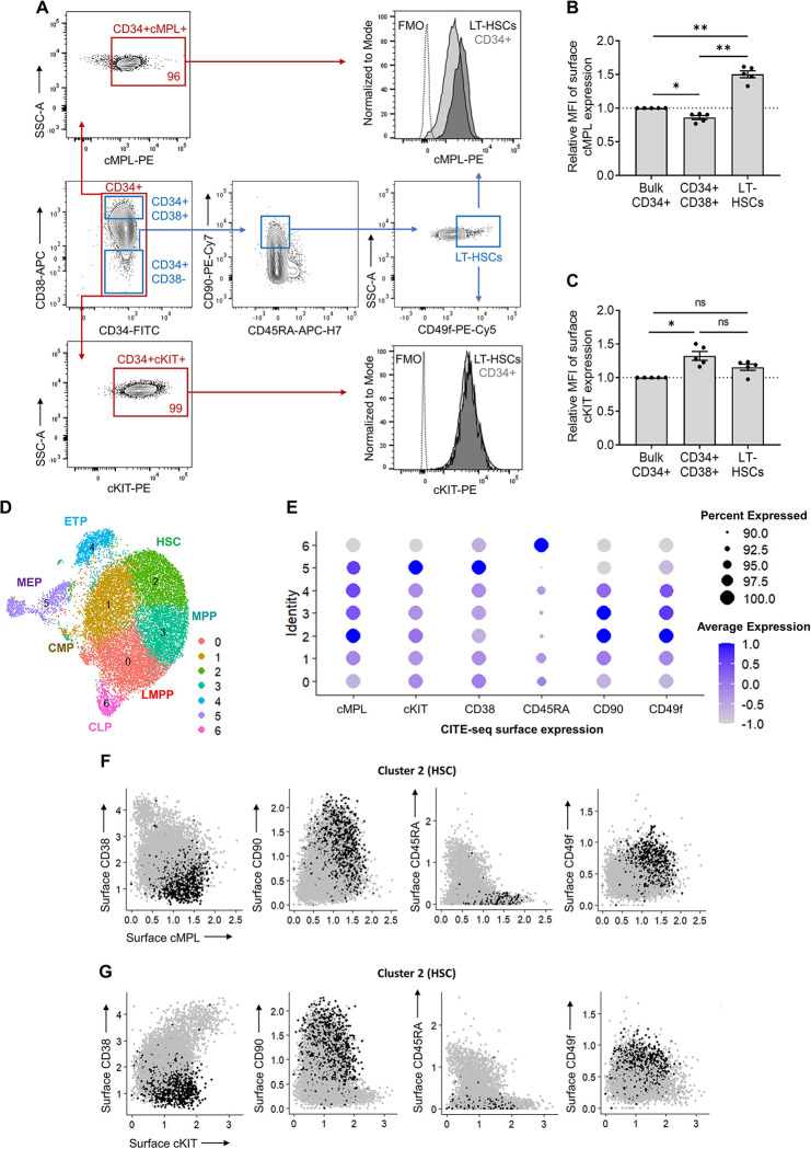
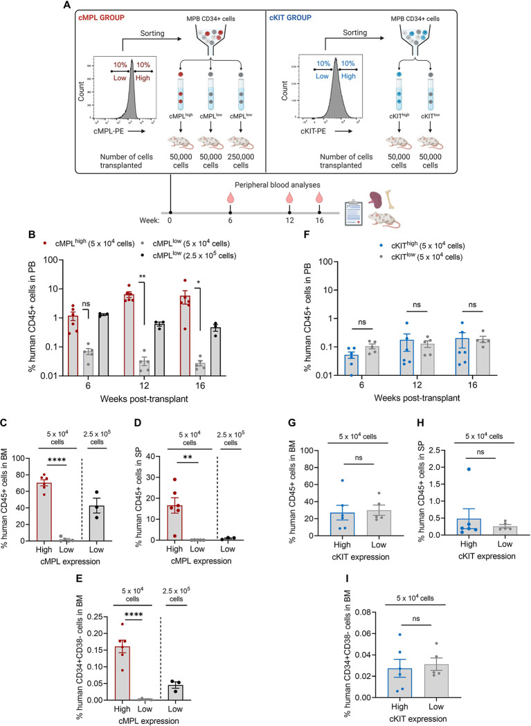
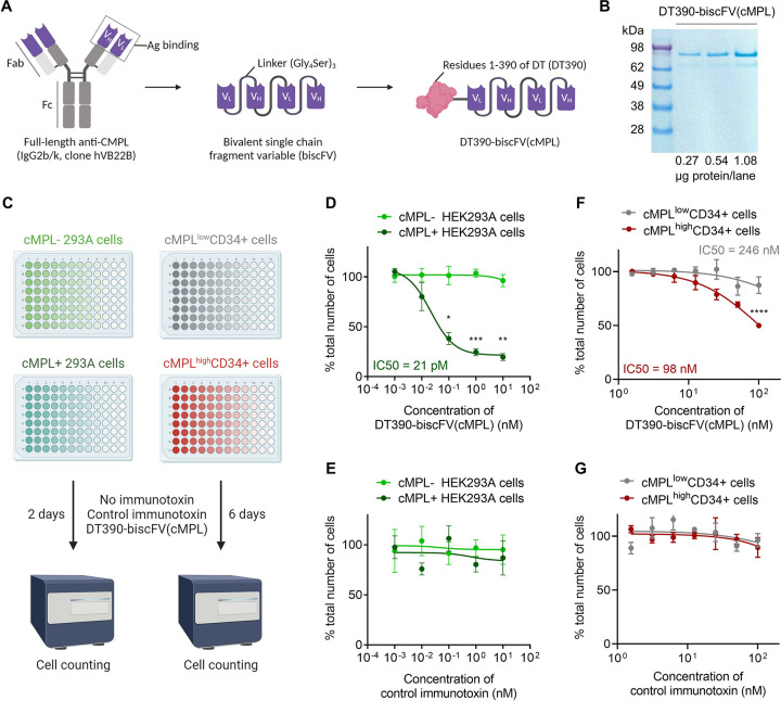
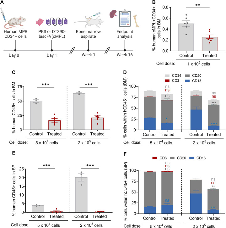
The transplantation of gene-modified autologous hematopoietic stem and progenitor cells (HSPCs) offers a promising therapeutic approach for hematological and immunological disorders. However, this strategy is often limited by the toxicities associated with traditional conditioning regimens. Antibody-based conditioning strategies targeting cKIT and CD45 antigens have shown potential in mitigating these toxicities, but their long-term safety and efficacy in clinical settings require further validation. In this study, we investigate the thrombopoietin (TPO) receptor, cMPL, as a novel target for conditioning protocols. We demonstrate that high surface expression of cMPL is a hallmark feature of long-term repopulating hematopoietic stem cells (LT-HSCs) within the adult human CD34+ HSPC subset. Targeting the cMPL receptor facilitates the separation of human LT-HSCs from mature progenitors, a delineation not achievable with cKIT. Leveraging this finding, we developed a cMPL-targeting immunotoxin, demonstrating its ability to selectively deplete host cMPLhigh LT-HSCs with a favorable safety profile and rapid clearance within 24 hours post-infusion in rhesus macaques. These findings present significant potential to advance our understanding of human hematopoiesis and enhance the therapeutic outcomes of ex vivo autologous HSPC gene therapies.


Conflict of interest statement

Declaration of Interests D.A, D.M., Z.W. and A.L. are inventors on the patent of DT390-biscFV(cMPL) (E-Numbers: E-188-2021-0). The other authors declare no competing financial interests.

Figures

Figure 1 |
Figure 1 |. High surface expression of cMPL enriches phenotypically and transcriptionally defined human HSCs.
(A) Flow cytometry gating strategy employed to resolve distinct populations within the CD34+ cell fraction of human mobilized peripheral blood (MPB). Initial gating on CD34+ cells (red gate) allowed for the subsequent separation into progenitors (CD34+CD38+) and LT-HSCs (CD34+CD38−CD90+CD45RA−CD49f+) (blue gates). The expression profiles of cMPL and cKit receptors were then evaluated within these subsets. The histograms on the right display representative mean fluorescence intensity (MFI) for cMPL and cKIT, illustrating their expression levels within the LT-HSC and CD34+ cell populations. Fluorescence minus one (FMO) is included as a control. (B, C) Quantitation of cMPL and cKIT surface expression in identified populations by MFI, normalized against the expression within total CD34+ cells (n = 5 independent donors). (D) Uniform Manifold Approximation and Projection (UMAP) clustering from CITE-seq data of human MPB CD34+ cells, identifying seven distinct HSPC clusters: lymphoid-primed multipotent progenitors (LMPP, cluster 0), common myeloid progenitors (CMP, cluster 1), hematopoietic stem cells (HSC, cluster 2), multipotent progenitors (MPP, cluster 3), early T-lineage progenitors (ETP, cluster 4), megakaryocytic-erythroid progenitors (MEP, cluster 5) and common lymphoid progenitors (CLP, cluster 6). (E) Dot plot visualization of protein surface expression from CITE-seq, with dot size reflecting cell expression percentage and color intensity denoting scaled average expression level across cluster types. (F, G) Feature plots from Seurat analysis correlating cMPL (F) and cKIT (G) expression with established HSC phenotypes within the transcriptionally defined HSC cluster 2. Datapoints in panels (B) and (C) represent the mean ± standard error of the mean (SEM), with statistical significance assessed via one-way ANOVA with Tukey correction. Notations of statistical significance are as follows: ns; not significant, * p ≤ 0.05, ** p ≤ 0.01.
Figure 2 |
Figure 2 |. The cMPL receptor serves as a marker for human long-term repopulating HSCs.
(A) Schematic of the experimental procedure: Human CD34+ cells from mobilized peripheral blood (MPB) were sorted into high and low subsets based on surface expression of cMPL and cKIT, followed by transplantation into NBSGW mice to assess hematopoietic reconstitution (n = 3–5 mice per group). (B-E) Comparative analysis of human cell engraftment in the peripheral blood (PB) (B), bone marrow (BM) (C), and spleen (SP) (D) of NBSGW mice after transplantation with human cMPLhighCD34+ or cMPLlowCD34+ cells, with panel (E) depicting the engraftment of HSC-enriched populations (CD34+CD38-) within the BM. (F-I) Comparative analysis of human cell engraftment in the PB (F), BM (G), and SP (H) of NBSGW mice after transplantation with human cKIThigh or cKITlow CD34+ cells, with panel (I) depicting the engraftment of HSC-enriched populations (CD34+CD38-) within the BM. Datapoints represent the mean ± standard error of the mean (SEM), with statistical significance assessed via two-sided unpaired t-tests. Notations of statistical significance are as follows: ns, not significant, * p ≤ 0.05, ** p ≤ 0.01, **** p ≤ 0.0001.
Figure 3 |
Figure 3 |. Significant enrichment of human LT-HSCs within cMPLhighCD34+ cells measured by a limiting-dilution secondary transplantation assay.
(A) Schematic of the experimental procedure: Human CD45+ cells, harvested from the bone marrow (BM) of primary recipient mice from both cMPLhigh and cMPLlow groups, underwent a secondary transplantation into NBSGW immunodeficient mice. The cells were transplanted at a limiting dilution after conditioning with a low busulfan dose, and BM engraftment levels were assessed 16 weeks following the transplantation (n = 4–15 mice per group). (B) Quantitative analysis of human CD45+ cell populations within the BM after secondary transplantation of cells derived from both cMPLhigh and cMPLlow groups. The dashed line represents the threshold (0.01%) above which the presence of human CD45+ cells, encompassing both myeloid (CD13+) and lymphoid (CD20+) lineages, is considered indicative of successful secondary engraftment. (C) Semilogarithmic representation of LT-HSC frequencies within the human CD45+ population post-secondary transplant, comparing the cMPLhigh and cMPLlow groups. The solid lines represent the best-fit linear regression for each group, while dotted lines indicate the 95% confidence intervals. (D) Quantitation of LT-HSC frequencies and absolute numbers derived from secondary transplantation assays conducted at limiting dilutions. LT-HSC counts within hCD45+ cells (column 8) were determined by multiplying LT-HSC frequencies (column 7) by the average pooled numbers of human CD45+ cells harvested from the primary mice’s limbs, including 4 hindlimb and 2 pelvic bones. Frequencies of LT-HSCs within human CD34+ cells (column 9) were established by dividing the LT-HSC numbers (column 8) by the initial quantity of hCD34+ cells transplanted into each primary mouse (column 2). The associated p-value was calculated using extreme limiting dilution statistics. Datapoints in panel (B), represent the mean ± standard error of the mean (SEM).
Figure 4 |
Figure 4 |. DT390-biscFV(cMPL) immunotoxin impairs the proliferation of cMPL-expressing human cell lines and HSPCs in vitro.
(A) Diagram of DT390-biscFV(cMPL) depicting the configuration of variable light (VL) and variable heavy (VH) chain sequences from the cMPL-specific monoclonal antibody hVB22B. These sequences are joined by (Gly4Ser)3 linkers, with the truncated diphtheria toxin (DT390) attached at the N-terminus of the biscFV(cMPL) to facilitate targeted cytotoxicity. (B) SDS-PAGE analysis of DT390-biscFV(cMPL) (molecular weight = 97 kDa). Lane 1: protein marker; Lanes 2 – 4: 0.27, 0.54 and 1.08 μg of DT390-biscFV(cMPL), respectively. (C) Schematic of the experimental procedure: In vitro cytotoxicity assay was performed using human cMPL-expressing HEK293A cells, control (cMPL−) HEK293A cells, human cMPLhigh mobilized peripheral blood (MPB) CD34+ cells or human cMPLlow MPB CD34+ cells. Cells were treated with DT390-biscFV(cMPL) or a non-specific control immunotoxin for 2 days (HEK293A cells) or 6 days (CD34+ cells). Cellular growth was assessed by automated counting, with values normalized to untreated control cultures. (D, E) Response of HEK293A cells to DT390-biscFV(cMPL) (D) or control immunotoxin (E) following a 2-day culture (n = 3 independent experiments). (F, G) Response of human MPB CD34+ cells to DT390-biscFV(cMPL) (F) or control immunotoxin (G) after a 6-day culture (n = 3 independent donors). Datapoints represent the mean ± standard error of the mean (SEM), with statistical significance assessed via two-sided unpaired t-tests. Notations of statistical significance are as follows: * p ≤ 0.05, ** p ≤ 0.01, *** p ≤ 0.001, **** p ≤ 0.0001.
Figure 5 |
Figure 5 |. DT390-biscFV(cMPL) depletes human cMPL+ HSPCs in a xenograft mouse model.
(A) Schematic of the experimental procedure: NBSGW mice were transplanted with human mobilized peripheral blood (MPB) CD34+ cells, followed by a single maximum tolerated dose of 1.2 mg/kg DT390-biscFv(cMPL) or PBS administered one day post-transplantation. Murine bone marrow (BM) was initially assessed at 1 week post-transplantation (cell dose: 1 × 10⁶), and subsequently both murine BM and spleen (SP) were evaluated at 16 weeks post-transplantation (cell dose: 5 × 10⁴ or 2 × 10⁵). (B) Percentage of human cMPL+CD34+ cells within the murine BM one week after administration of PBS (control) or DT390-biscFV(cMPL) (treated) (n = 5–6 mice per group). (C, D) Percentage of human CD45+ cells (C) and their lineage distribution (D) within the murine BM at the 16-week endpoint analysis for control and treated groups across both transplantation cell doses (n = 3–5 mice per group). (E, F) Percentage of human CD45+ cells (E) and their lineage distribution (F) within the murine SP at the 16-week endpoint analysis for control and treated groups across both transplantation cell doses (n = 3–5 mice per group). Datapoints represent the mean ± standard error of the mean (SEM), with statistical significance assessed via two-sided unpaired t-tests (panels B, C and E) or two-way ANOVA with Sidak multiple comparisons tests (panels D, F). Notations of statistical significance are as follows: ns, not significant, ** p ≤ 0.01, *** p ≤ 0.001, **** p ≤ 0.0001.
Figure 6 |
Figure 6 |. DT390-biscFV(cMPL) preferentially depletes LT-HSCs in rhesus macaques.
(A) Relative mean fluorescence intensity (MFI) of cMPL expression on the cell surface within specified hematopoietic populations, normalized to the MFI of cMPL on bulk CD34+ cells (n = 4 animals per group). (B) Schematic of the experimental procedure: a single intravenous infusion of DT390-biscFv(cMPL) was administered at dosages of 0.2, 0.4, 0.6, or 0.8 mg/kg to four separate rhesus macaques. Post-administration, the impact on hematopoietic cell populations was monitored through serial sampling of bone marrow (BM) and peripheral blood. (C) Representative flow cytometry plots exemplifying the cMPLhighCD34+ cell populations within BM aspirates pre-treatment (baseline) and four days after a 0.6 mg/kg dose of DT390-biscFv(cMPL). (D-F) Frequency of primitive hematopoietic subsets in BM samples collected pre-treatment and on days 4, 18, 56, and 365 following treatment (n = 1 animal per DT390-biscFV(cMPL) dose tested). Hematopoietic populations displayed include cMPLhighCD34+ cells (D), LT-HSCs (characterized as CD34+CD38−CD90+CD45RA−CD49f+ cells) (E), and bulk CD34+ cells (F). Datapoints in panels A, and D-F represent the mean ± standard error of the mean (SEM). In panel (A), statistical significance was assessed via one-way ANOVA with Tukey correction. Notations of statistical significance are as follows: ns, not significant, * p ≤ 0.05, ** p ≤ 0.01.
Figure 7 |
Figure 7 |. Safety and Pharmacokinetics Profile of DT390-biscFV(cMPL) in rhesus macaques.
(A) Pharmacokinetic profile: The graph illustrates the time-dependent serum concentration of DT390-biscFV(cMPL) post-administration (n = 3 technical replicates at each timepoint). The dotted line represents the assay’s sensitivity threshold at 15 ng/mL. Note that the drug’s concentration falls below the detectable limit within 24 hours, highlighting its short systemic half-life. Legend in panel A applies to all panels. (B) Temporal changes in platelet counts following DT390-biscFV(cMPL) administration. (C) Luminex assay data indicating the serum thrombopoietin (TPO) levels post-treatment (n = 2 technical replicates at each timepoint). (D) Hemoglobin levels after treatment. (E) Neutrophil trajectory after treatment. (F) Lymphocyte counts after treatment. (G) Alanine transaminase (ALT) levels measured after treatment with DT390-biscFV(cMPL) to assess hepatic function and potential drug-induced hepatotoxicity. (H) Creatinine levels measured after treatment with DT390-biscFV(cMPL) to evaluate renal function and potential drug-induced nephrotoxicity. The dotted lines depicted in panels B and D-H represent the established normal range for each blood parameter measured. Datapoints in panels A and C represent the mean ± standard error of the mean (SEM).

References

    1. Morgan R.A., Gray D., Lomova A. & Kohn D.B. Hematopoietic Stem Cell Gene Therapy: Progress and Lessons Learned. Cell Stem Cell 21, 574–590 (2017). - PMC - PubMed
    1. Tucci F., Scaramuzza S., Aiuti A. & Mortellaro A. Update on Clinical Ex Vivo Hematopoietic Stem Cell Gene Therapy for Inherited Monogenic Diseases. Mol Ther 29, 489–504 (2021). - PMC - PubMed
    1. Dunbar C.E., et al. Gene therapy comes of age. Science 359(2018). - PubMed
    1. Bernardo M.E. & Aiuti A. The role of conditioning in hematopoietic stem cell gene therapy. Hum Gene Ther (2016). - PubMed
    1. Shaw P., Shizuru J., Hoenig M., Veys P. & Iewp E. Conditioning Perspectives for Primary Immunodeficiency Stem Cell Transplants. Front Pediatr 7, 434 (2019). - PMC - PubMed

Publication types