[Preprint]. 2024 Feb 29:2024.02.28.582502.
doi: 10.1101/2024.02.28.582502.

Magnesium Transporter MgtA revealed as a Dimeric P-type ATPase

Affiliations

Magnesium Transporter MgtA revealed as a Dimeric P-type ATPase

Rilee Zeinert et al. bioRxiv. .

Update in

  • P-type ATPase magnesium transporter MgtA acts as a dimer.
    Zeinert R, Zhou F, Franco P, Zöller J, Madni ZK, Lessen H, Aravind L, Langer JD, Sodt AJ, Storz G, Matthies D. Zeinert R, et al. Nat Struct Mol Biol. 2025 Jun 23. doi: 10.1038/s41594-025-01593-7. Online ahead of print. Nat Struct Mol Biol. 2025. PMID: 40550995

Abstract

Magnesium (Mg2+) uptake systems are present in all domains of life given the vital role of this ion. Bacteria acquire Mg2+ via conserved Mg2+ channels and transporters. The transporters are required for growth when Mg2+ is limiting or during bacterial pathogenesis, but, despite their significance, there are no known structures for these transporters. Here we report the first structure of the Mg2+ transporter MgtA solved by single particle cryo-electron microscopy (cryo-EM). Using mild membrane extraction, we obtained high resolution structures of both a homodimeric form (2.9 Å), the first for a P-type ATPase, and a monomeric form (3.6 Å). Each monomer unit of MgtA displays a structural architecture that is similar to other P-type ATPases with a transmembrane domain and two soluble domains. The dimer interface consists of contacts between residues in adjacent soluble nucleotide binding and phosphotransfer regions of the haloacid dehalogenase (HAD) domain. We suggest oligomerization is a conserved structural feature of the diverse family of P-type ATPase transporters. The ATP binding site and conformational dynamics upon nucleotide binding to MgtA were characterized using a combination of cryo-EM, molecular dynamics simulations, hydrogen-deuterium exchange mass spectrometry, and mutagenesis. Our structure also revealed a Mg2+ ion in the transmembrane segments, which, when combined with sequence conservation and mutagenesis studies, allowed us to propose a model for Mg2+ transport across the lipid bilayer. Finally, our work revealed the N-terminal domain structure and cytoplasmic Mg2+ binding sites, which have implications for related P-type ATPases defective in human disease.

Keywords: P-type ATPase; cryo-EM; dimer; ion translocation; magnesium; membrane protein; oligomer; transport.


Conflict of interest statement

Competing interests The authors declare no competing interests.

Figures

Fig 1
Fig 1. Cryo-EM of magnesium transporter MgtA reveals a high-resolution dimer and a monomeric structure.
a, Schematic representation of MgtA/B based on P-type ATPase structural homology showing ten conserved transmembrane helices colored in purple (1-10), the actuator (A) domain in orchid, the phosphorylation (P) subdomain in light blue, the nucleotide (N) binding or CAP subdomain in light green, and the predicted unstructured N-terminal tail in orange. The A domain is split into two regions a/b. The b segment of the A domain is comprised of a Double Stranded beta-Helix fold (DSβH). The soluble P subdomain is a noncontiguous segment comprised of two regions a/b that house the key catalytic residues required for phosphorylation. The N subdomain binds the nucleotide and aids in catalysis. The P and N subdomains together comprise the Haloacid dehalogenase (HAD) domain, b, Representative micrograph of purified Escherichia coli MgtA. c-d, Representative final 2D class averages for the ~200 kDa dimer (c) and the ~100 kDa monomer (d), respectively, with a box size of 384 pixels (approximately 319 Å). e-f, Cryo-EM reconstruction of the MgtA dimer (e, also see Extended Data Fig. 9a and Extended Data Movie 1) and of the monomer (f, also see Extended Data Fig. 9b and Extended Data Movie 2). Cryo-EM maps in e-f are colored as in a. A transparent gray map at a much lower threshold indicates the detergent micelle and the density for the more flexible loops and the A domain in the monomer map.
Fig 2
Fig 2. The dimer interface is formed by both hydrophobic and polar interactions.
a, Side view of the overall dimer structure with the left monomer in gray and the right monomer colored as in Fig. 1. A close-up view of the extensive dimer interface between the two N and P subdomains with sidechain residues displaying charge interactions across the dimer interface. Rotation of the structure highlights hydrophobic interactions at the dimer interface. Molecular dynamic simulations (see Extended Data Movies 5 and 6) show consistent interactions across the dimer interface between K382-E582 (64%), Q380-Q380 (87%), and K548-E549 (88%) shown in bold. b, Co-purification of two differentially tagged MgtA derivatives, one tagged with His6 and the other tagged with the larger SPA tag as shown in the top schematic reveals copurification of the two proteins (right elution panel). Proteins were visualized by Western blot analysis using MgtA antibodies.
Fig 3
Fig 3. The nucleotide binding pocket of MgtA is accessible in the dimeric state.
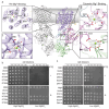
a, Side view of the MgtA dimer with the left monomer in gray and the right monomer colored as in Fig. 1, highlighting the ATP molecule represented in spheres located in between the soluble A domain and P and N subdomains and the dephosphorylation TGES loop in yellow. b, A close-up view of the ATP binding site highlighting residues in close proximity to the Mg-ATP molecule which is shown in a ball and stick representation and the cryo-EM map density in gray mesh. Residues from the TGES loop involved in dephosphorylation and located in the A domain are colored yellow. c, Distances from ATP to amino acids, water molecules and Mg2+ ion in the nucleotide binding pocket of MgtA from E. coli, determined using LIGPLOT+ . d, MgtAD373N is unable to completement a Mg2+-auxotrophic E. coli strain indicating this mutant transporter does not translocate Mg2+ ions. D373 is the residue that is being phosphorylated upon ATP hydrolysis. MgtAE215A is only partially able to complement upon overexpression. E215 is part of the TGES loop involved in dephosphorylation. Overnight cultures were serial diluted and spotted onto LB agar plates supplemented with high (100 mM) or low (1 mM) MgSO4 with (+) and without (−) 0.1 mM IPTG for induction and grown at 37°C (also see Extended Data Fig. 21).
Fig 4
Fig 4. Mg2+ binds sites near the middle of the transmembrane domain and between the cytosolic A domain and N and P subdomains.
a, Side view of the overall dimeric structure with the left monomer in gray and the right monomer colored as in Fig. 1 with close-up views of the resolved Mg2+ ions and the nearby residues and proximal resolved water molecules (small red spheres). Site I is located within the TM α-helices 5, 7, and 8. Site II is comprised of residues from the A domain and P subdomain. Site III is comprised of residues from the A domain and N subdomain. b, MgtAE331A (located in TM4) and MgtAD780A (located in TM7) are unable to complement a Mg2+-auxotrophic E. coli strain. c, MgtAD441A (site III) is only partially able to complement the Mg2+-auxotrophic E. coli strain upon overexpression. Complementation assays in b and c were carried out as for Fig. 3d (also see Extended Data Fig. 23).
Fig 5
Fig 5. Summary of MgtA structural insights.
a, Proposed Mg2+ion path based on structural, conservation analysis, and MD simulation data. b, Overview of putative regulatory features of MgtA including the dimer interface, cytoplasmic Mg2+ binding sites, N-terminal domain, lipid environment, and small protein binding. Green circles represent Mg2+ ions. Color scheme is the same as in Fig. 1.

Similar articles

References

    1. Moomaw A. S. & Maguire M. E. The unique nature of Mg2+ channels. Physiology 23, 275–285 (2008). - PMC - PubMed
    1. Franken G. A. C., Huynen M. A., Martínez-Cruz L. A., Bindels R. J. M. & de Baaij J. H. F. Structural and functional comparison of magnesium transporters throughout evolution. Cell. Mol. Life Sci. 79, 418 (2022). - PMC - PubMed
    1. Jin F., Huang Y. & Hattori M. Recent advances in the structural biology of Mg2+ channels and transporters. J. Mol. Biol. 434, 167729 (2022). - PubMed
    1. García V. et al. Genome-wide analysis of fitness-factors in uropathogenic Escherichia coli during growth in laboratory media and during urinary tract infections. Microb. Genom. 7, 000719 (2021). - PMC - PubMed
    1. Papp-Wallace K. M. et al. The CorA Mg2+ channel is required for the virulence of Salmonella enterica serovar Typhimurium. J. Bacteriol. 190, 6517–6523 (2008). - PMC - PubMed

Publication types