[Preprint]. 2025 Apr 2:2024.02.28.582599.
doi: 10.1101/2024.02.28.582599.

Phosphate-binding pocket on cyclin B governs CDK substrate phosphorylation and mitotic timing

Affiliations

Phosphate-binding pocket on cyclin B governs CDK substrate phosphorylation and mitotic timing

Henry Y Ng et al. bioRxiv. .

Update in

Abstract

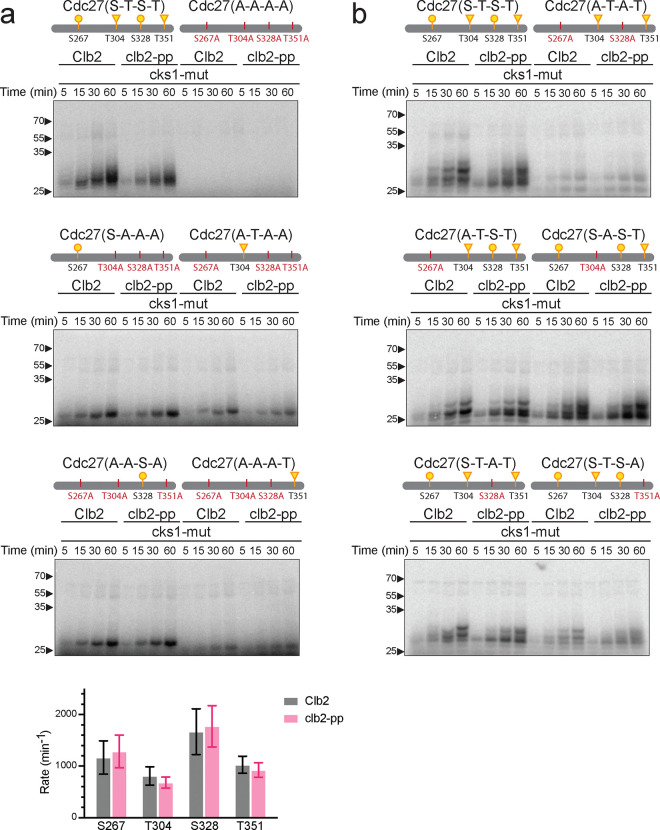
Cell cycle progression is governed by complexes of the cyclin-dependent kinases (CDKs) and their regulatory subunits cyclin and Cks1. CDKs phosphorylate hundreds of substrates, often at multiple sites. Multisite phosphorylation depends on Cks1, which binds initial priming phosphorylation sites to promote secondary phosphorylation at other sites. Here, we describe a similar role for a recently discovered phosphate-binding pocket (PP) on B-type cyclins. Mutation of the PP in Clb2, the major mitotic cyclin of budding yeast, alters bud morphology and delays the onset of anaphase. Mutation of the PP reduces multi-site phosphorylation of CDK substrates in vitro, including the Cdc16 and Cdc27 subunits of the anaphase-promoting complex/cyclosome and the Bud6 and Spa2 subunits of the polarisome. We conclude that the cyclin PP, like Cks1, controls the pattern of multisite phosphorylation on CDK substrates, thereby helping to establish the robust timing of cell-cycle events.


Conflict of interest statement

Competing interests The authors declare no competing interests.

Figures

Fig. 1:
Fig. 1:. Mutation of the conserved phosphate-binding pocket in Clb2 causes a growth defect in the absence of CLB1 and CLB3.
a, Aligned amino acid sequences of B-type cyclins, with cyan indicating the three basic residues that constitute the phosphate-binding pocket. These residues are not conserved in A-type cyclins. b, (left) Growth curves of indicated yeast strains, with OD600 recorded every 15 min. Data represent mean ± standard deviation (SD) of four independent biological replicates. (right) Growth rates calculated by fitting OD600/min to a logistic growth model. Data represent mean ± SD of four independent biological replicates. Statistical significance was determined using one-way analysis of variance (ANOVA) (****p < 0.0001). c, Representative differential interference contrast (DIC) microscopy images of the indicated yeast strains at mid-log phase in rich (YPD) media at 30°C. Scale bar is 10 μm.
Fig. 2:
Fig. 2:. The clb2-pp mutation causes a mitotic delay.
a, Single-cell measurements of time from SPB separation to spindle elongation in asynchronous wild-type (WT) or clb2-pp cells carrying mCherry-labeled Spc42. Each data point represents a single cell and the bars indicate median ± SD. The sample size (n) is noted at right. See Supplementary Fig. 2a for representative images. b, Western blots of Myc-tagged securin after release from a G1 arrest in the indicated yeast strains. Alpha factor was added back at the 60-min time point. GAPDH loading control on lower blot. Representative of 10 independent experiments. c, Western blots of Myc-tagged Ndd1 after release from a G1 arrest in the indicated yeast strains. These strains carry a second copy of Ndd1-Myc under the control of a GAL promoter, and experiments were performed in galactose media. Alpha factor was added back at the 60-min time point. GAPDH loading control on lower blot. Representative of 2 independent experiments. d, Same as b with swe1Δ. Representative of 2 independent experiments. e, Same as b with mad2Δ. Representative of 2 independent experiments. f, Same as b with apc1-Δloop. Representative of 2 independent experiments.
Fig. 3:
Fig. 3:. Phospho-pocket mutations reduce phosphorylation of APC/C subunits in vitro.
a, APC/C was immunoprecipitated from yeast lysate and treated with wild-type or clb2-pp Clb2-Cdk1 plus mutant Cks1 and radiolabeled ATP. Reaction products were analyzed by SDS-PAGE and autoradiography (lane 1: kinase only control; lane 2: no kinase control; lane 3–6: wild-type Clb2; lane 7–10: Clb2-pp). APC/C subunits were identified based on previous studies. b, 2.5 μM purified Cdc16 fragment (aa 31–180) was incubated with 150 nM wild-type or clb2-pp Clb2-Cdk1 plus wild-type or mutant Cks1 and radiolabeled ATP. Diagrams at top indicate suboptimal (S/T*-P; yellow) CDK consensus sites (S: circle; T: triangle) in the fragment (see Supplementary Fig. 4 for complete sequences). Representative of 10 independent experiments. c, Same as b with 2.5 μM purified Cdc27 fragment (aa 241–360) and 100 nM Clb2-Cdk1. Representative of 8 independent experiments. Coomassie Blue-stained gels for b and c are found in Supplementary Fig. 6.
Fig. 4:
Fig. 4:. Phospho-pocket mutations reduce phosphorylation of CDK consensus sites on Cdc27.
a, b, 2.5 μM of the indicated Cdc27 mutant fragments were incubated with 100 nM wild-type or clb2-pp Clb2-Cdk1 plus mutant Cks1 and radiolabeled ATP. Reaction products were analyzed by Phos-tag SDS-PAGE and autoradiography. Diagrams at top indicate suboptimal (S/T*-P; yellow) CDK consensus sites (S: circle; T: triangle) in the tested fragment (see Supplementary Fig. 4 for complete sequences). Representative of 4 independent experiments. Coomassie Blue-stained gels in Supplementary Fig. 7. For the graph at bottom of panel a, phosphate incorporation into the single site mutants over time was used to calculate the mean phosphorylation rate (± SD) in 4 independent experiments.
Fig. 5:
Fig. 5:. Phospho-pocket mutations reduce phosphorylation of polarisome subunits.
2.5 μM of purified Bud6 fragment (aa 126–360) or 5 μM of purified Spa2 fragment (aa 524–670) was incubated with 100 nM wild-type or clb2-pp Clb2-Cdk1 plus wild-type or mutant Cks1 and radiolabeled ATP. Diagrams at top indicate optimal (S/T*-P-x-K/R; green) and suboptimal (S/T*-P; yellow) CDK consensus sites (S: circle; T: triangle) in the tested fragment (see Supplementary Fig. 4 for complete sequences). Representative of 3 independent experiments. Coomassie Blue-stained gels in Supplementary Fig. 8.
Fig. 6:
Fig. 6:. Stoichiometry of phosphorylation at specific sites on CDK substrates.
The indicated five substrates (10 μM) were incubated for 60 min in 55 μl reaction volumes containing 200 nM wild-type or clb2-pp Clb2-Cdk1 plus 200 nM mutant Cks1. Samples were trypsinized, and seven aliquots from each reaction were subjected to LC-MS/MS. High-confidence phosphopeptides (2% ion intensity, AScore 15) were tabulated with the PTM Profiles function in PEAKS software, which provides the total peak areas for modified peptides and the corresponding unmodified peptide (Supplementary Table 3). The fraction of modified peptide is plotted here (mean +/− SD, where n is 4 to 7 replicates with quantifiable peak areas). Diagrams at top indicate optimal (S/T*-P-x-K/R; green) and suboptimal (S/T*-P; yellow) CDK consensus sites. Blue indicates non-consensus sites lacking a proline at +1 but containing a basic (K/R) residue at +3 or +4; other non-consensus sites are colored gray (see Supplementary Fig. 4 for complete sequences). A small number of CDK consensus sites were not confidently quantified and are shown as vertical marks (S95 in Cdc16, S283 in Bud6, S544 and T549 in Spa2).

References

    1. Ubersax J. A. et al. Targets of the cyclin-dependent kinase Cdk1. Nature 425, 859–864 (2003). - PubMed
    1. Holt L. J. et al. Global analysis of Cdk1 substrate phosphorylation sites provides insights into evolution. Science (1979) 325, 1682–1686 (2009). - PMC - PubMed
    1. Loog M. & Morgan D. O. Cyclin specificity in the phosphorylation of cyclin-dependent kinase substrates. Nature 434, 104–108 (2005). - PubMed
    1. Coudreuse D. & Nurse P. Driving the cell cycle with a minimal CDK control network. Nature 468, 1074–9 (2010). - PubMed
    1. Swaffer M. P., Jones A. W., Flynn H. R., Snijders A. P. & Nurse P. CDK Substrate Phosphorylation and Ordering the Cell Cycle. Cell 167, 1750–1761.e16 (2016). - PMC - PubMed

Publication types