[Preprint]. 2024 Feb 28:2024.02.24.581789.
doi: 10.1101/2024.02.24.581789.

Efficient and reproducible generation of human iPSC-derived cardiomyocytes using a stirred bioreactor

Affiliations

Efficient and reproducible generation of human iPSC-derived cardiomyocytes using a stirred bioreactor

Maksymilian Prondzynski et al. bioRxiv. .

Update in

Abstract

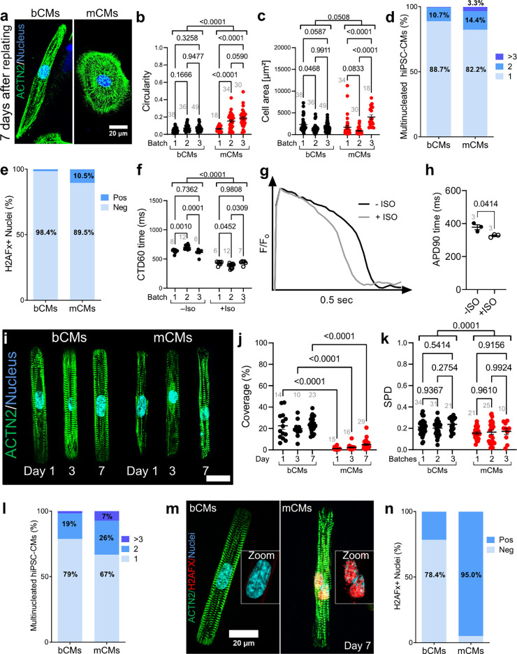
In the last decade human iPSC-derived cardiomyocytes (hiPSC-CMs) proved to be valuable for cardiac disease modeling and cardiac regeneration, yet challenges with scale, quality, inter-batch consistency, and cryopreservation remain, reducing experimental reproducibility and limiting clinical translation. Here, we report a robust cardiac differentiation protocol that uses Wnt modulation and a stirred suspension bioreactor to produce on average 124 million hiPSC-CMs with >90% purity using a variety of hiPSC lines (19 differentiations; 10 iPSC lines). After controlled freeze and thaw, bioreactor-derived CMs (bCMs) showed high viability (>90%), interbatch reproducibility in cellular morphology, function, drug response and ventricular identity, which was further supported by single cell transcriptomes. bCMs on microcontact printed substrates revealed a higher degree of sarcomere maturation and viability during long-term culture compared to monolayer-derived CMs (mCMs). Moreover, functional investigation of bCMs in 3D engineered heart tissues showed earlier and stronger force production during long-term culture, and robust pacing capture up to 4 Hz when compared to mCMs. bCMs derived from this differentiation protocol will expand the applications of hiPSC-CMs by providing a reproducible, scalable, and resource efficient method to generate cardiac cells with well-characterized structural and functional properties superior to standard mCMs.


Conflict of interest statement

Disclosures The authors have no competing interests to disclose.

Figures

Fig. 1:
Fig. 1:. Optimized stirred bioreactor cardiac differentiation protocol.
(a) Schematic representation of the optimized bioreactor cardiac differentiation protocol. (b-c) Characteristics of successful bioreactor cardiomyocyte differentiations. Runs were categorized as failed when they yielded < 90% hiPSC-CMs. hiPSC-CMs with frequency of pluripotency marker SSEA4 by flow cytometry (b) or out of range mean EB diameter (c) had higher likelihood of failure. n=19–25 differentiations. EB diameter analysis at day 0. (d) Spontaneous beating frequency of bioreactor- and monolayer-derived hiPSC-CMs at day 15 (n=number of differentiations; number of EBs or wells: Bioreactor (n=3; 71); Monolayer (n=3; 46). (e-f) hiPSC-CM yield (e) and purity (f) at day 15 of bioreactor or monolayer-directed cardiac differentiation (bioreactor, n=19 differentiations; monolayer, n=7 differentiations). Percentage of cells positive for cardiomyocyte marker cTnT was measured by flow cytometry. (g) Timeline of bioreactor and monolayer cardiac differentiation monitored by GFP fluorescence from a TNNI1-GFP iPSC line (bar: 200 μm). (h-j). qRT-PCR analysis of marker gene expression during bioreactor and monolayer cardiac differentiation. Values are expressed as fold-change compared to bioreactor day 5. ACTN2 (h) marks cardiomyocytes, COL1A1 (i) and COL3A1 (j) mark fibroblasts. n=number of differentiations; number of replicates: Bioreactor (n=2–3; 6–11); Monolayer (n=2–3; 6–11). (k) Quantification of viable hiPSC-CMs after cryo-recovery from bioreactor (n=10) and monolayer (n=3) differentiations. Data are expressed as mean ± SEM. b-f,k, Welch’s unpaired t-test. h-j, two-way ANOVA with Holm-Šidákś post-test. MCB, Master cell bank; human induced pluripotent stem cells,hiPSCs; EB, embryoid body; cardiac troponin T, cTnT; CM, cardiomyocyte; BF, bright field.
Fig. 2:
Fig. 2:. ScRNAseq reveals higher cardiomyocyte content and degree of cellular specification in bioreactor-derived hiPSC-CMs.
(a) ScRNA-seq UMAP clustering of monolayer (ML) and bioreactor-derived hiPSC-CMs (bCMs) showing 11 clusters and their assigned cell types (right). (b) Stacked bar graph showing cellular composition of ML (left) and bCMs (right) expressed in % as parts of whole. Clusters are divided in cardiomyocytes (top) and non-CMs (bottom), whereby the color coding is the same as in panel A, and the order of clusters and the corresponding % are indicated in the table below (b´). (c) Violin-plot showing the relative expression of a subset of cardiac and non-cardiac marker genes (y-axis) across all clusters for bCMs (grey) and mCMs (red) (x-axis). Bioreactor-derived cardiomyocytes, bCMs; Monolayer-derived cardiomyocytes, mCMs; Noncardiomyocytes, non-CMs.
Fig. 3:
Fig. 3:. Comparison of bCMs and mCMs on 2D platforms.
(a-e) Morphological characteristics of unpatterned hiPSC-CMs. Cryo-recovered bCMs and mCMs were cultured for 7 days on unpatterned Geltrex-coated dishes. Cells were stained for sarcomere Z-line marker ACTN2. Representative images (a) illustrate elongated shape of bCMs compared to mCMs. Bar, 20 μm. Circularity (b) and cell area (c) were quantified from 3 independent differentiation batches of bCMs and mCMs. Grey numbers indicate cells analyzed. Two-way ANOVA with Šidákś post-test. (d) Nucleation of unpatterned bCMs (n=178) and mCMs (n=118) after 7 days in culture. Chi-squared p<0.0001. (e) Level of DNA damage response in unpatterned bCMs and mCMs. bCMs (n=63) and mCMs (n=95) were stained for H2AFX, which accumulates as a reaction to DNA double strand breaks. Chi-squared p<0.0001. (f) bCM calcium transients. Unpattered cryo-recovered bCMs were loaded with Ca2+ sensitive dye Fluo-4 and Ca2+ transients were optically recorded during 1 Hz pacing after 4 days of culture. mCMs could not be paced so were excluded. Calcium transient duration at 60% recovery (CTD60) was measured from 3 independent differentiation batches without (filled circles; n=26 wells) and with (open circles; n=13 wells) 1 μM isoproterenol (ISO) after 4 days in culture. Grey numbers indicate number of wells analyzed. Two-way ANOVA with Šidákś post-test. (g-h) bCM action potentials. Unpattered cryo-recovered bCMs were loaded with voltage sensitive dye Fluovolt and optically recorded during 1 Hz pacing after 7 days in culture. g, Representative action potential traces for bCMs without and with (grey line) 1 μM isoproterenol (ISO). h, Quantification of action potential duration at 90% recovery (APD90) for bCMs without (n=3 wells) and with (n=3 wells) 1 μM isoproterenol (ISO). Three separate wells were analyzed per group. Data are expressed as mean ± SEM, paired t-test. (i-k) Characterization of micropatterned bCMs and mCMs. Cryo-recovered cells were plated on single cell extracellular matrix rectangular islands. Samples were fixed at stained after 1, 3, and 7 days in culture. i, Representative images. Bar, 20 μm. (j) Unbiased quantification of bCMs and mCMs coverage of single cell islands. Grey numbers indicate 10x fields analyzed. Two-way ANOVA with Šidákś post-test. (k) Unbiased analysis of sarcomere packing density (SPD) of micropatterned bCMs and mCMs 7 days after replating. Grey numbers indicate cells analyzed. Two-way ANOVA with Šidákś post-test. (l) Nucleation of micropatterned bCMs (n=192) and mCMs (n=60) after 7 days in culture. Chi-squared p<0.0001. (m) Level of DNA damage response in micropatterned bCMs and mCMs. Cells were stained for ACTN2 and H2AFX. m, representative images. Scale bar, 20 μm. (n) Quantification of H2AFx positive and negative nuclei in bCMs (n=51) and mCMs (n=20). Chi-squared p<0.0001. Data are expressed as mean ± SEM. Bioreactor-derived cardiomyocytes, bCMs; Monolayer-derived cardiomyocytes, mCMs; Isoproterenol, ISO.
Fig. 4:
Fig. 4:. Comparison of 3D engineered heart tissues (EHTs) constructed with bCM or mCMs.
(a) Representative images of EHTs after 29 days in culture (Scale bar, 1 mm). (b-e) Spontaneously beating EHTs assembled from cryo-recovered bCMs or mCMs were recorded in culture medium at 37°C from day 5 to day 32. Analyses of baseline frequency (b), force (c), time to 50% contraction (C50; d) and 90% relaxation (R90; e) showed greater force generation in bCM EHTs. Two-way ANOVA with Šidákś post-test of pooled bCMs and mCMs values for each timepoint. (f-h) Analysis of EHTs in Tyrode solution (f-h) without pacing (0 Hz) or with 1–3 Hz pacing. f, EHT beat frequency in response to pacing. Only EHTs captured by pacing are shown. The percent of EHTs captured at each pacing rate is indicated. Two-way ANOVA with Šidákś post-test. (i) Histological characterization of bCM and mCM EHTs. Cryosections of EHTs after 34 days in culture were stained for sarcomere Z-line protein ACTN2. Representative cryosections (i) showed higher cellularity and greater sarcomere content and organization in bCM EHTs. Sarcomere length (j) was quantified from 33 (bCM) or 35 (mCM) regions of interest from 2 (bCM) or 3 (mCM) EHTs. Data are expressed as mean ± SEM. Mann-Whitney test. Bioreactor-derived cardiomyocytes, bCMs; Monolayer-derived cardiomyocytes, mCMs.

References

    1. Hamad S. et al. Generation of human induced pluripotent stem cell-derived cardiomyocytes in 2D monolayer and scalable 3D suspension bioreactor cultures with reduced batch-to-batch variations. Theranostics 9, 7222–7238 (2019). - PMC - PubMed
    1. Lian X. et al. Directed cardiomyocyte differentiation from human pluripotent stem cells by modulating Wnt/β-catenin signaling under fully defined conditions. Nat. Protoc. 8, 162–175 (2013). - PMC - PubMed
    1. Chen V. C. et al. Development of a scalable suspension culture for cardiac differentiation from human pluripotent stem cells. Stem Cell Res. 15, 365–375 (2015). - PMC - PubMed
    1. Kempf H., Kropp C., Olmer R., Martin U. & Zweigerdt R. Cardiac differentiation of human pluripotent stem cells in scalable suspension culture. Nat. Protoc. 10, 1345–1361 (2015). - PubMed
    1. Breckwoldt K. et al. Differentiation of cardiomyocytes and generation of human engineered heart tissue. Nat. Protoc. 12, 1177–1197 (2017). - PubMed

Publication types