[Preprint]. 2024 Feb 29:2024.02.29.582594.
doi: 10.1101/2024.02.29.582594.

Hachiman is a genome integrity sensor

Affiliations

Hachiman is a genome integrity sensor

Owen T Tuck et al. bioRxiv. .

Update in

Abstract

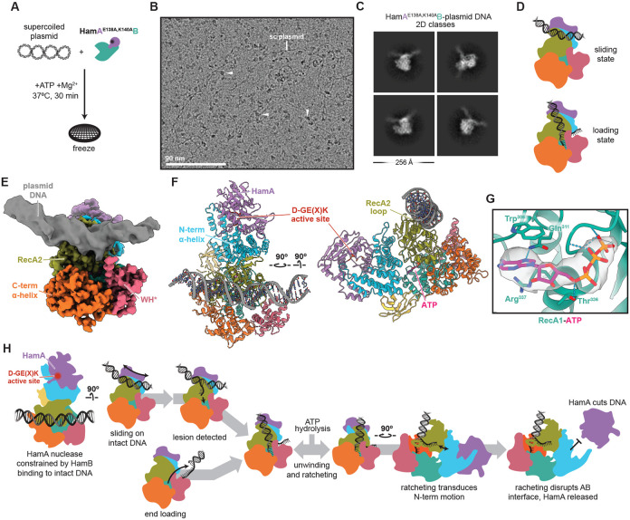
Hachiman is a broad-spectrum antiphage defense system of unknown function. We show here that Hachiman comprises a heterodimeric nuclease-helicase complex, HamAB. HamA, previously a protein of unknown function, is the effector nuclease. HamB is the sensor helicase. HamB constrains HamA activity during surveillance of intact dsDNA. When the HamAB complex detects DNA damage, HamB helicase activity liberates HamA, unleashing nuclease activity. Hachiman activation degrades all DNA in the cell, creating 'phantom' cells devoid of both phage and host DNA. We demonstrate Hachiman activation in the absence of phage by treatment with DNA-damaging agents, suggesting that Hachiman responds to aberrant DNA states. Phylogenetic similarities between the Hachiman helicase and eukaryotic enzymes suggest this bacterial immune system has been repurposed for diverse functions across all domains of life.


Conflict of interest statement

Competing interest statement J.A.D. is a co-founder of Caribou Biosciences, Editas Medicine, Intellia Therapeutics, Mammoth Biosciences and Scribe Therapeutics, and a director of Altos, Johnson & Johnson and Tempus. J.A.D. is a scientific advisor to Caribou Biosciences, Intellia Therapeutics, Mammoth Biosciences, Inari, Scribe Therapeutics, Felix Biosciences and Algen. J.A.D. also serves as Chief Science Advisor to Sixth Street and a Scientific Advisory Board member at The Column Group. J.A.D. conducts academic research projects sponsored by Roche and Apple Tree Partners. J.P. has an equity interest in Linnaeus Bioscience Incorporated and receives income. The terms of this arrangement have been reviewed and approved by the University of California, San Diego, in accordance with its conflict-of-interest policies.

Figures

Figure 1 ∣
Figure 1 ∣. Hachiman is a two-component defense system that protects against diverse bacteriophages.
(A) Overview of SF1/SF2 helicase-containing phage defense systems found in RefSeq genomes in the DefenseFinder database. (B) Phylogenetic tree of core helicase domains of 329 helicases from defense systems from (A) and representative SF1/SF2 helicases. Helicase superfamily is provided in the outer track (SF1 in black, SF2 in tan) and representative families demarcated in gray clades with labels. Defense system-associated helicases are colored as shown in (A). Details on tree construction and sequence alignment provided in methods. (C) Hachiman loci from E. coli strains ECOR04, ECOR28 and ECOR31 tested in this study. HamA genes are shown in purple and HamB genes shown in green. Additional defense systems identified in PADLOC are shown in blue, integrases in yellow and tRNA genes in red. All other genes are shown in gray. (D) Overview of phage-defense assays. Native Hachiman loci are cloned under an anhydrotetracycline (aTc)-inducible promoter, pTet, and monitored for protection against diverse phages. (E) Representative plaque assays for ECOR31 HamAB against sensitive phages EdH4 and T4 as well as resistant phage T5. (F) Comparison of different Hachiman loci against 12 diverse phages representing 12 unique phage genera. Plaque assays without EOP reductions, but a measurable difference in plaque size are denoted with an asterisk. (G) Protection against phage EdH4 is complete at low MOI, but insufficient at high MOI. For panels (E-G), Hachiman is induced at 20nM aTc and dCas13d targeting RFP is provided as a negative control. All assays consist of 3 biological replicates.
Figure 2 ∣
Figure 2 ∣. Structural basis of Hachiman complexation and identification of the HamA active site.
(A) Cryo-EM density of the E. coli ECOR31 apo HamAB complex. The sharpened map is colored, while the unsharpened map is overlaid and transparent. (B) Orthogonal views of the HamAB structure, with domains colored according to the key above. Walker motifs are annotated in the HamB RecA1 and RecA2 domains. (C) Overview of the HamA-HamB NAH interface, with surfaces involved in the interaction shown. (D-F) Detail of three subregions, HamA- (D), HamA102-117 (E), and HamA159-199 (F), contributing to the AB interface. Residues contributing to hydrogen bonding interactions are shown as sticks and are labeled with colors corresponding to the key above each view and in (B). (G) Sequence logo resulting from alignment of HamA DUF1837 ORFs. The ECOR31 HamA sequence and corresponding position is shown below each residue logo. (I) Plaque assays demonstrating the ability of HamAB and various mutants to confer defense against phage EdH4. Individual data points of three independent biological replications are shown along with the mean and standard deviation. The (−) symbol indicates a reduction in plaque size.
Figure 3 ∣
Figure 3 ∣. HamAB is a DNA nuclease/helicase that degrades plasmids in vitro.
(A) Malachite green ATPase assays of HamB against a panel of nucleic acid substrates. Individual data points of three independent biological replicates and the mean and standard deviation are shown. (B-E) HamB DNA unwinding assays on substrates with a 15 bp duplex and a 15 nt 3′ OH (B), forked 15 nt OH (C), 15 nt 5′ OH (D), and no overhang (E). DNA substrates are labeled with 5′ FAM. Gels are representative of three independent replicates. (F) Normalized percent unwinding of DNA substrates with 15 bp (circles), 25 bp (squares), and 50 bp (triangles) duplex lengths, all labeled with 5′ FAM and with a 15 nt 3′ OH. Individual data points shown are quantifications of replications of unwinding assays in the format of B-E normalized against basal unwinding (see Methods). (G) In vitro plasmid clearance assay after 90 min at 37°C with ATP using MBP-HamA, HamB, HamAB, and HamA*B visualized on a 0.75% agarose gel. (H) Time course of HamAB plasmid clearance with addition of ATP or SSB visualized on a native agarose gel. (I) Time course assay as in J with mutant HamA*B. (J) ATPase activity of HamB, HamAB, and HamA*B with or without supercoiled plasmid substrates. Individual data points of three independent biological replicates and the mean and standard deviation are shown. (K) Cartoon summarizing in vitro activities of HamB. (L) Cartoon depicting a model for HamAB-mediated plasmid degradation.
Figure 4 ∣
Figure 4 ∣. Structural basis of HamB-DNA binding and helicase ratcheting.
(A) Cryo-EM density of the 2.8 Å HamB-DNA density. The sharpened map is colored according to domain, while the unsharpened map is overlaid and transparent. (B) Orthogonal views of the 2.8 Å HamB-DNA structure. (C) Detail of the 3’ end of the DNA buried within the DNA entry site of HamB. Hydrogen bonds and contributing residues are shown with a dashed line. (D) Detail of the DNA duplex-interacting RecA2 loop. (E) Left, superimposed conformers of HamB-DNA viewed from the DNA side, with conformation 1 (2.8 Å) colored teal and conformation 2 (2.9 Å) colored burgundy. Right, conformations 1 and 2 viewed from the NAH side and transparent, with vectors colored according to domain representing motion between the two conformations. Vectors are scaled 2x and are calculated using modevectors. (F) Representative disruption of the predicted AB interface between the two HamB conformations. AB interactions disrupted by HamB motion are shown and labeled. (G) Native PAGE of reactions of the HamAB complex with the DNA where ratcheting was observed in cryo-EM. ATP and DNA appear to dissociate the AB complex. (H) Model for HamB signal transduction to the NAH and concomitant release of HamA.
Figure 5 ∣
Figure 5 ∣. Hachiman defends against bacteriophage and prevents lysis by nonspecific DNA clearance.
(A) Wildtype HamAB, HamAB* (HamABD431A) or HamA*B (HamAE138A,K140AB) expressing E. coli either uninfected (−EdH4) or 60 minutes post-infection by phage EdH4 (+EdH4). Scale bar = 3 μm. Cell membranes were stained with FM4-64 (red) and DNA was stained with DAPI (cyan). (B) Quantification of percentage of cells with completely degraded chromosomes. The following lists the sample size, in number of cells, for each condition: WT HamAB, −EdH4 = 188 cells; WT HamAB, +EdH4 = 211 cells; HamAB*, −EdH4 = 185 cells; HamAB*, +EdH4 = 213 cells; HamA*B, −EdH4 = 171 cells; HamA*B, +EdH4 = 180 cells. (C) Examples of DNA morphologies in uninfected and EdH4-infected cells expressing HamAB* or HamA*B. Scale bar = 1 μm. Cell membranes were stained with FM4-64 (red) and DNA was stained with DAPI (cyan).
Figure 6 ∣
Figure 6 ∣. DNA damage activates Hachiman.
(A-C) Cell growth of E. coli expressing wildtype HamAB, HamA*B and HamAB* at 20nM aTc at 20nM aTc in the absence or presence of subinhibitory and inhibitory concentrations of nalidixic acid (A), novobiocin (B) or gentamycin (C). Growth curves are colored according to condition. See Fig. S6 for complete minimum inhibitory concentration (MIC) determinations. All measurements were performed in biological triplicate.
Figure 7 ∣
Figure 7 ∣. Hachiman scans intact dsDNA.
(A) HamA*B-plasmid +ATP specimen preparation. (B) Representative motion-corrected, dose-weighted cryo-EM micrograph from the HamA*B-plasmid DNA dataset. Plasmid DNA and bound particles are indicated with white arrows. (C) Representative 2D classes of particles bound to plasmid DNA. (D) Cartoon depicting the scanning state resolved here and comparison with the loading state resolved in the HamB-DNA dataset. (E) Composite cryo-EM density colored according to domain. Protein regions are from the 3.2 Å deepEMhancer-sharpened map, while DNA is from the 3.2 Å sharp map masked and B-factor refined to display helical features. (F) Orthogonal views of the HamA*B-plasmid DNA structure. The DNA sequence is unknown. (G) Detail of ATP in HamB, with residues and hydrogen bonds shown. The density is masked to ATP. (H) Proposed mechanism of Hachiman immunity.

References

    1. Singleton M. R., Dillingham M. S. & Wigley D. B. Structure and 1. Singleton M. R., Dillingham M. S. & Wigley D. B. Structure and mechanism of helicases and nucleic acid translocases. Annu. Rev. Biochem. 76, 23–50 (2007). - PubMed
    1. Pyle A. M. Translocation and unwinding mechanisms of RNA and DNA helicases. Annu. Rev. Biophys. 37, 317–336 (2008). - PubMed
    1. Bleichert F., Botchan M. R. & Berger J. M. Mechanisms for initiating cellular DNA replication. Science 355, (2017). - PubMed
    1. Fuller-Pace F. V. DExD/H box RNA helicases: multifunctional proteins with important roles in transcriptional regulation. Nucleic Acids Res. 34, 4206–4215 (2006). - PMC - PubMed
    1. Jarmoskaite I. & Russell R. RNA helicase proteins as chaperones and remodelers. Annu. Rev. Biochem. 83, 697–725 (2014). - PMC - PubMed

Publication types