[Preprint]. 2024 Feb 23:rs.3.rs-3970621.
doi: 10.21203/rs.3.rs-3970621/v1.

Complement propagates visual system pathology following traumatic brain injury

Affiliations

Complement propagates visual system pathology following traumatic brain injury

Davis Borucki et al. Res Sq. .

Update in

Abstract

Background: Traumatic brain injury (TBI) is associated with the development of visual system disorders. Visual deficits can present with delay and worsen over time, and may be associated with an ongoing neuroinflammatory response that is known to occur after TBI. Complement activation is strongly associated with the neuroinflammatory response after TBI, but whether it contributes to vision loss after TBI is unexplored.

Methods: Acute and chronic neuroinflammatory changes within the dorsal lateral geniculate nucleus (dLGN) and retina were investigated subsequent to murine controlled unilateral cortical impact. Neuroinflammatory and histopathological data were interpreted in the context of behavioral and visual function data. To investigate the role of complement, cohorts were treated after TBI with the complement inhibitor, CR2-Crry.

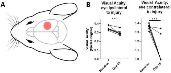
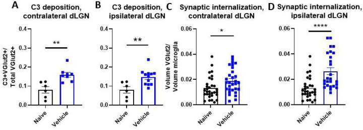
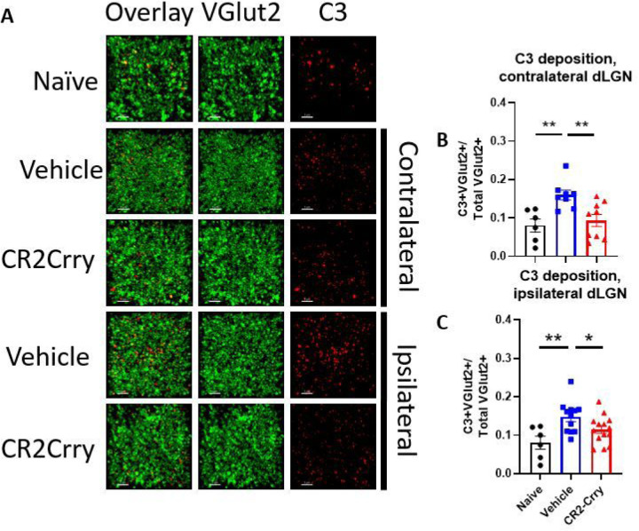
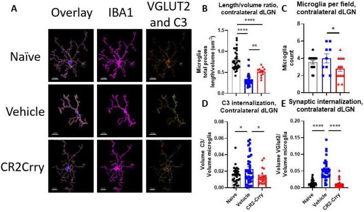
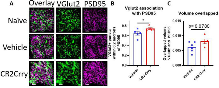
Results: At 3 days after TBI, complement C3 was deposited on retinogeniculate synapses in the dLGN both ipsilateral and contralateral to the lesion, which was reduced in CR2-Crry treated animals. This was associated with microglia morphological changes in both the ipsilateral and contralateral dLGN, with a more amoeboid phenotype in vehicle compared to CR2-Crry treated animals. Microglia in vehicle treated animals also had a greater internalized VGlut2+ synaptic volume after TBI compared to CR2-Crry treated animals. Microglia morphological changes seen acutely persisted for at least 49 days after injury. Complement inhibition also reduced microglial synaptic internalization in the contralateral dLGN and increased the association between VGLUT2 and PSD95 puncta, indicating preservation of intact synapses. Unexpectedly, there were no changes in the thickness of the inner retina, retinal nerve fiber layer or retinal ganglion layer. Pathologies were accompanied by reduced visual acuity at subacute and chronic time points after TBI, with improvement seen in CR2-Crry treated animals.

Conclusion: TBI induces complement activation within the dLGN and promotes microglial activation and synaptic internalization. Complement inhibition after TBI in a clinically relevant paradigm reduces complement activation, maintains a more surveillance-like microglia phenotype, and preserves synaptic density within the dLGN. Together, the data indicate that complement plays a key role in the development of visual deficits after TBI via complement-dependent microglial phagocytosis of synapses within the dLGN.


Conflict of interest statement

Competing interests The authors have no competing interests.

Figures

Figure 1
Figure 1
Controlled cortical impact produces a significant decline in visual function. (a) Schematic of injury location. Mice receive a craniotomy and a single, direct, right-sided impact on the dura (B) Mice experience deficits in visual acuity as measured by the optomotor response after injury, with most mice experiencing a complete loss in contralateral vision, and a significant decrease in ipsilateral visual acuity. b, paired t-test. ***p<0.001.
Figure 2
Figure 2
Controlled cortical impact results in increased complement deposition on retinogeniculate synapses and increased synaptic internalization by microglia (a-b) Proportion of synapses colocalizing with C3 in the dLGN contralateral (a) and ipsilateral (b) to injury. (c-d) Microglial internalization of excitatory retinogeniculate synaptic marker VGLUT2 in contralateral (c) and ipsilateral (d) dLGNs. a-d t-test, *p<0.05, **p<0.01. Error bars = mean ± s.e.m.
Figure 3
Figure 3
Complement deposits on synapses in the dLGN, both ipsilateral and contralateral to injury, and is reduced by complement inhibition. (a) Representative images showing C3 (red) and VGLUT2 (green) colocalization within the dLGN. Scale bar = 5 μm. (b) Proportion of synapses colocalizing with C3 in the contralateral dLGN. (c) Proportion of synapses colocalizing with C3 in the ipsilateral dLGN. , t-test, *p<0.05, **p<0.01. Error bars = mean ± s.e.m.
Figure 4
Figure 4
Microglia internalize VGLUT2+ synapses and C3 after injury and display morphological features indicative of activation in both dLGN, and these changes are reduced by complement inhibition. (a) Representative microglial reconstructions with internalized VGLUT2 (green) and C3 (red), and microglial morphology (IBA1, magenta). Scale bar = 10 μm. (b) Microglial filament length to volume ratio, (c) microglial internalization of C3, and (d) internalization of VGLUT2 in the contralateral dLGN. (e) Microglia filament length to volume ratio, (f) microglial internalization of C3, and (g) microglial internalization of VGLUT2 in the ipsilateral dLGN. c, f: t-test. b, d, e, and f: one-way ANOVA with Tukey correction for multiple comparisons. *p<0.05, **p<0.01, ***p<0.001, ****p<0.0001. Error bars = mean ± s.e.m.
Figure 5
Figure 5
Injury site-targeted complement inhibition prevents the formation of visual deficits in the ipsilateral eye after controlled cortical impact. (a) Treatment paradigm. CR2-Crry (16 mg/kg) or saline was administered via i.v. tail vein injection one hour after CCI. Behavioral testing was performed at the timepoints indicated. (b) Acute inhibition of complement activation preserved visual function in the ipsilateral eye, as measured by the optomotor response. (c) Animals almost always step away from the cliff when it is in their ipsilateral field of vision. (d) Both vehicle and CR2-Crry-treated animals step toward the cliff or safe sides with equal frequency when the cliff is in their contralateral field of vision. (e) Acute inhibition of complement activation significantly reduced time to find the escape hole in the Barnes maze. b-e one-way ANOVA with Tukey correction for multiple comparisons. *p<0.05, **p<0.01, ***p<0.001. Error bars = mean ± s.e.m.
Figure 6
Figure 6
Microglia in the contralateral dLGN still internalize VGLUT2+ synapses and C3 and display morphological features indicative of activation along with increased numbers at a chronic timepoint. (a) Representative microglial reconstructions with internalized VGLUT2 (green) and C3 (red), and microglial morphology (IBA1, magenta). Scale bar = 10 μm. (b) Microglia filament length to volume ratio in the contralateral dLGN. (c) Microglia count per 63x high power field. (d) Microglial internalization of C3 and (d) microglial internalization of VGLUT2 in the contralateral dLGN. B, d-e one-way ANOVA with Tukey correction for multiple comparisons. c t-test. *p<0.05, **p<0.01, ***p<0.001, ****p<0.0001. Error bars = mean ± s.e.m.
Figure 7
Figure 7
Synaptic density in the contralateral dLGN is preserved by complement inhibition. (a) Representative images showing VGLUT2 (green) and PSD95 (magenta) colocalization within the contralateral dLGN. Scale bar = 5 μm. (b) Proportion of VGLUT2 puncta within 0.2 μm of PSD95 puncta. (c) Total volume overlapped between VGLUT2 and PSD95. t-test, *p<0.05. Error bars = mean ± s.e.m.
Figure 8
Figure 8
Controlled cortical impact does not cause appreciable changes in retinal thickness, but does result in increased complement staining in the retina contralateral to injury. (a) There is an increase in C3 staining intensity in the contralateral eye, both in the inner retina and full thickness retina, that is reduced by complement inhibition. There are no significant differences detected in the ipsilateral retina. (b) There are no significant changes in the thickness of the retinal nerve fiber layer of either eye at 42 days after injury, as measured by optical coherence tomography in living animals.

References

    1. Hac NEF, Gold DR. Neuro-Visual and Vestibular Manifestations of Concussion and Mild TBI. Curr Neurol Neurosci. 2022;22(3):219–28. - PubMed
    1. Bell CA, Grossman SN, Balcer LJ, Galetta SL. Vision as a piece of the head trauma puzzle. Eye (Basingstoke). 2023. - PMC - PubMed
    1. Goodrich GL, Flyg HM, Kirby JE, Chang C-Y, Martinsen GL. Mechanisms of TBI and Visual Consequences in Military and Veteran Populations. Optometry Vis Sci. 2013;90(2). - PubMed
    1. Chen B, Zhang H, Zhai Q, Li H, Wang C, Wang Y. Traumatic optic neuropathy: a review of current studies. Neurosurg Rev. 2022;45(3):1895–913. - PubMed
    1. Kaur P, Sharma S. Recent Advances in Pathophysiology of Traumatic Brain Injury. Curr Neuropharmacol. 2018;16(8):1224–38. - PMC - PubMed

Publication types

LinkOut - more resources