

[Importance of cone beam computed tomography in the recognition of the trajectory and anatomical variants of the mandibular canal. A review of the literature]
- PMID: 38464412
- PMCID: PMC10919828
- DOI: 10.21142/2523-2754-0901-2021-046
[Importance of cone beam computed tomography in the recognition of the trajectory and anatomical variants of the mandibular canal. A review of the literature]
Abstract
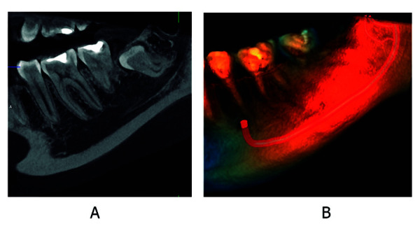
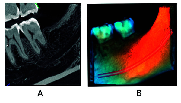
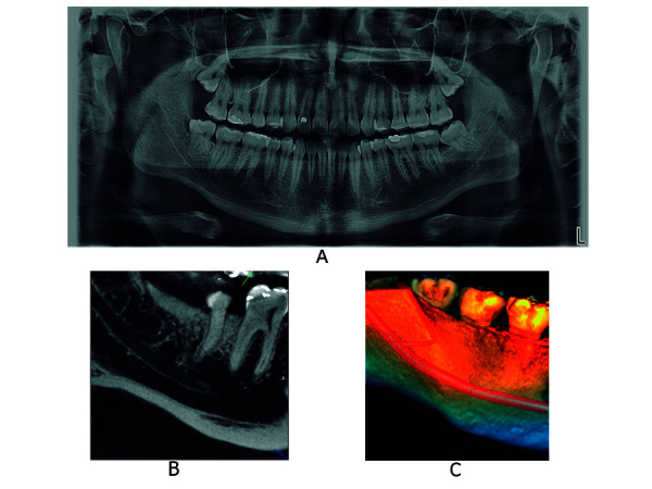
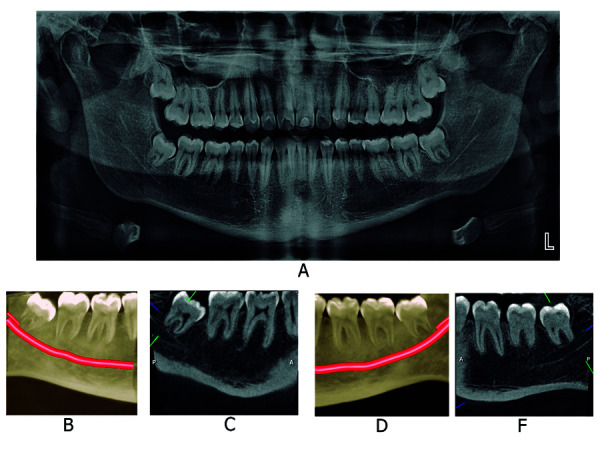
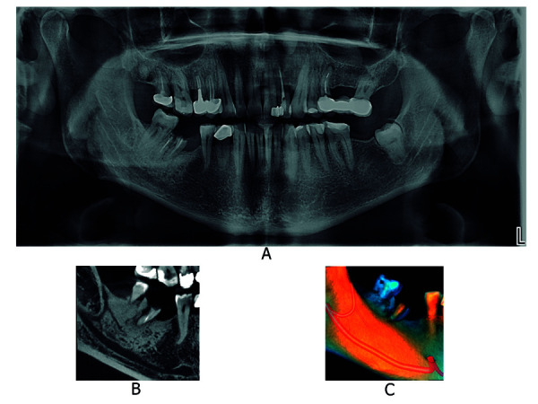
The objective of this study was to provide an updated review of the literature on the importance of the use of cone beam computed tomography (CBCT) in the recognition of the trajectory and variants of the mandibular canal (MCV).CBCT allows obtaining high quality images and visualization with an accuracy of approximately 94%, compared to 53% with periapical intraoral radiography (RIP) and 17% with panoramic extraoral radiography (REP), making CBCT an important diagnostic tool.The incidences of MCV in CBCT studies were between 1.3% and 69%, with differences between patients of different ethnic origins and within the same ethnic population, and in the types and configurations of MCV within each ethnic group. The studies available in the literature provide a histological description of the content of MCV. The presence of nerve and artery bundles of different calibers suggests that patients present clinical symptoms only if the neurovascular bundle reaches a certain size and number of fascicles. This review provides a description of the different classifications available and updated with CBCT.
El objetivo de este estudio fue realizar una revisión actualizada de la literatura sobre la importancia del uso de la tomografía computarizada de haz cónico (TCHC) en el reconocimiento de la trayectoria y las variantes del canal mandibular (VCM), ya que esta permite obtener imágenes de alta calidad, con una exactitud de 94%, aproximadamente, mientras que la radiografía intraoral periapical (RIP) tiene un 53% y la radiografía extraoral panorámica (REP) presenta un 17% de exactitud. Las incidencias de las variantes del canal mandibular en estudios realizados utilizando TCHC fueron entre un 1,3% y un 69%. Estas pueden diferir entre los pacientes de diferentes orígenes étnicos y, a su vez, dentro de la misma población étnica; además, hay grandes diferencias en los tipos y configuraciones de las VCM dentro de cada grupo étnico. Estudios realizados han demostrado histológicamente el contenido de las VCM; la presencia de haces de nervios y arterias de diferentes calibres sugieren también que los pacientes presentan síntomas clínicos solamente si el paquete neurovascular alcanza cierto tamaño y número de fascículos. En este estudio se describieron las diferentes clasificaciones realizadas y actualizadas con TCHC.
Keywords: bifid mandibular canal; cone beam computed tomography; mandibular canal; trifid mandibular canal.
Conflict of interest statement
Potenciales conflictos de interés: Los autores declaran no tener conflicto de intereses de ningún tipo
Figures
Similar articles
-
Bifid mandibular canal: confirmation of limited cone beam CT findings by gross anatomical and histological investigations.Dentomaxillofac Radiol. 2012 Sep;41(6):460-5. doi: 10.1259/dmfr/60245722. Epub 2011 Nov 24. Dentomaxillofac Radiol. 2012. PMID: 22116121 Free PMC article.
-
Assessment of bifid and trifid mandibular canals using cone-beam computed tomography.Imaging Sci Dent. 2014 Sep;44(3):229-36. doi: 10.5624/isd.2014.44.3.229. Epub 2014 Sep 17. Imaging Sci Dent. 2014. PMID: 25279344 Free PMC article.
-
Comparative analysis of mandibular anatomical variations between panoramic radiography and cone beam computed tomography.Oral Maxillofac Surg. 2014 Dec;18(4):419-24. doi: 10.1007/s10006-013-0428-z. Epub 2013 Aug 24. Oral Maxillofac Surg. 2014. PMID: 23975215
-
What are the retromolar and bifid/trifid mandibular canals as seen on cone-beam computed tomography? Revisiting classic gross anatomy of the inferior alveolar nerve and correcting terminology.Surg Radiol Anat. 2022 Jan;44(1):147-156. doi: 10.1007/s00276-021-02862-y. Epub 2021 Dec 2. Surg Radiol Anat. 2022. PMID: 34854962 Review.
-
Anatomical variations of the mandibular canal and their clinical implications in dental practice: a literature review.Surg Radiol Anat. 2021 Aug;43(8):1259-1272. doi: 10.1007/s00276-021-02708-7. Epub 2021 Feb 25. Surg Radiol Anat. 2021. PMID: 33630105
References
-
- Quispe ML. Tomographic characteristics of the bifurcation of the mandibular canal. Rev Estomatol Herediana. 2016;26(3):122–131.
-
- Domínguez J, Ruge O, Aguilar G, Ñáñez Ó, Oliveros G. Cone beam computed tomographic analysis of the position and course of the mandibular canal. Rev Fac Odontol Univ Antioq. 2010;22(1):12–22.
-
- Parihar A, Warhekar SA, Gharote HP, Warhekar AM. Bifid mandibular canal An unusual presentation. J Indian Acad Oral Med Radiol. 2015;27:453–456. doi: 10.4103/0972-1363.170485. - DOI
-
- Ya-Qiong Z, Zh Ya-Ning, Deng-Gao L, Yuan M, Xu-Chen M. Bifid variations of the mandibular canal cone beam computed tomographic evaluation of 1000 Northern Chinese patients. Oral surgery, oral medicine, oral pathology and oral radiology. 2018;126(5):271–278. doi: 10.1016/j.oooo.2018.06.008. - DOI - PubMed
-
- Chávez-Lomeli ME, Mansilla-Lory J, Pompa JA, Kjaer I. The human mandibular canal arises from three separate canals innervating different tooth groups. Dent Res. 1996;75(8):1540–1544. - PubMed
Publication types
LinkOut - more resources
Full Text Sources
Miscellaneous