Review
. 2024 Mar 6:13:151-172.
doi: 10.2147/ITT.S381472. eCollection 2024.

Dermatomyositis: Practical Guidance and Unmet Needs

Affiliations
Review

Dermatomyositis: Practical Guidance and Unmet Needs

Lydia Cassard et al. Immunotargets Ther. .

Abstract

Dermatomyositis is a heterogeneous idiopathic inflammatory myopathy associated with various cutaneous manifestations and variable presence of myositis, interstitial lung disease, and other visceral organ involvement. An accurate diagnosis of dermatomyositis requires correlating clinical examination findings with serological and histological findings. Familiarity with pathognomonic and common cutaneous manifestations of dermatomyositis, which are highlighted here, can be especially helpful in making an accurate diagnosis. Additionally, evaluating patients for presence of myositis-specific autoantibodies can further support or refute a dermatomyositis diagnosis. When present, myositis-specific autoantibodies can also help guide workups for various dermatomyositis-associated manifestations, as each is associated with relatively distinct clinical characteristics. Evaluating patients for various systemic manifestations often relies on expert opinion recommendations; however, societal guideline statements concerning the evaluation of some manifestations have recently been described. Although malignancy-associated dermatomyositis is a well-accepted subtype, there is limited evidence to support extensive malignancy screening has a favorable benefit-risk ratio in most dermatomyositis patients. However, recent research has uncovered novel associations between dermatomyositis and malignancy, suggesting the possibility of identifying high-risk subsets of dermatomyositis patients in whom malignancy screening may have a high value. Treatment for dermatomyositis has remained largely unchanged over the past several decades. Although many dermatomyositis patients can be effectively treated with current options, either as monotherapy or with combination regimens, there is a need for more targeted and effective DM therapies, in general, and for MDA5(+) dermatomyositis-associated rapidly progressive interstitial lung disease. Fortunately, significant current and emerging research activities evaluating various novel medications for dermatomyositis provide hope for exciting future advances in patients with this intriguing immune-mediated disease.

Keywords: MDA-5; autoantibody; idiopathic inflammatory myopathy; malignancy; myositis; treatment.


Conflict of interest statement

APF: Investigator for Pfizer, Corbus, Mallinckrodt, Alexion, Priovant, and Novartis receives personal research support from Mallinckrodt and Novartis; honorarium from AbbVie, BMS, Biogen, Novartis, and UCB for consultation and advisory board participation; and honorarium from AbbVie, Novartis, BMS, Kyowa Kirin, and Mallinckrodt for teaching and speaking. The other authors have no conflicts of interest to declare for this work.

Figures

Figure 1
Figure 1
Pathognomonic Cutaneous Manifestations of Dermatomyositis. (A) A 69-year-old man with violaceous periorbital erythema and edema (heliotrope rash) related to TIF1γ(+) DM. He was subsequently diagnosed with diffuse large B-cell lymphoma. This patient provided written consent to use this image for publication. (B) A 60-year-old woman with TIF1γ autoantibodies and erythematous-to-violaceous papules overlying the interphalangeal joints of the hands (Gottron’s papules) related to TIF1γ(+) DM. (C) A 55-year-old woman with erythematous-to-violaceous patches on the knees (Gottron’s sign) related to TIF1γ(+) DM. (D) A 52-year-old woman with erythematous-to-violaceous patches on the elbows (Gottron’s sign) related to SAE(+) DM.
Figure 2
Figure 2
Common Cutaneous Manifestations of Dermatomyositis. (A) A 55-year-old woman with erythematous patches and plaques involving the upper back and shoulders (shawl sign) related to TIF1γ(+) DM; (B) A 54-year-old man with erythematous patches and plaques involving the lateral Hip (holster sign) related to DM. He declined MSA testing; (C) A 78-year-old man with erythematous patches involving the anterior neck and upper chest (V-neck erythema) related to TIF1γ(+) DM; (D) A 66-year-old woman with erythematous-to-violaceous patches and plaques with thick, psoriasiform scale and associated alopecia involving the scalp in relation to TIF1γ(+) DM.
Figure 3
Figure 3
Other Cutaneous Manifestations of Dermatomyositis. (A) A 68-year-old woman with long-standing DM and skin atrophy, telangiectasia, and dyspigmentation (poikiloderma) in a V-neck distribution; (B) A 54-year-old man with long-standing DM and firm, white nodules of calcinosis cutis (arrows) involving his scalp; (C) A 42-year-old woman with Jo1(+) Antisynthetase syndrome with significant fissuring and hyperkeratosis of her fingers (Mechanic’s hands); (D) A 68-year-old woman with TIF1γ autoantibodies with (arrows) dilated proximal nailfold capillary loops, capillary dropout, and focal cuticular hemorrhage related to TIF1γ(+) DM.
Figure 4
Figure 4
Characteristic Histologic Features of Lesional Dermatomyositis Skin Biopsies. (A) A photomicrograph (Hematoxylin and Eosin staining; 100X magnification) of a lesional punch biopsy from the upper back (shawl area) of a 55-year-old Caucasian woman with TIF1γ DM revealing vacuolar interface dermatitis affecting the epidermis, mild superficial dermal lymphocytic inflammation, and abundant mucin deposition throughout the reticular dermis denoted by areas of blue discoloration (arrows) between collagen bundles. (B) At higher power (Hematoxylin and Eosin staining; 400X magnification), dyskeratotic keratinocytes (the hallmark feature of interface dermatitis) are seen as cells along the basilar epidermis with pyknotic nuclei and condensed eosinophilic cytoplasm (white arrow) and eosinophilic globules just below the basilar epidermis (black arrow).
Figure 5
Figure 5
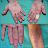
Characteristic Cutaneous Manifestations of MDA5-associated Dermatomyositis. (A) A 52-year-old man with MDA5(+) DM and deep, punched out ulcers involving the palmar and lateral aspects of numerous fingers. Note the associated violaceous discoloration suggestive of underlying vasculopathy. (B) A 42-year-old woman with MDA5(+) DM-associated palmar papules with central ivory-white discoloration and surrounding violaceous erythema near the creases of interphalangeal joints. (C) A 67-year-old man with MDA5(+) DM-associated crusted erosions and ulcers and violaceous erythema involving his elbow and forearm.
Figure 6
Figure 6
MDA5(+) Dermatomyositis-associated Rapidly Progressive Interstitial Lung Disease. A 58-year-old woman admitted with shortness-of-breath and a rash consisting of (A) crusted ulcerations involving the V-neck area and (B) violaceous-to-hyperpigmented papules overlying the interphalangeal joints of her hands. A lesional punch biopsy from the V-neck area revealed vacuolar interface dermatitis and (C) CT chest at baseline revealed patchy opacities throughout the lungs and focal areas of fibrosis. Despite aggressive treatment her pulmonary opacities and fibrotic CT changes progressed and pulmonary function decreased over (D) 2 months and (E) 7 months and she eventually expired from her disease.

References

    1. Khoo T, Lilleker JB, Thong BY-H, Leclair V, Lamb JA, Chinoy H. Epidemiology of the idiopathic inflammatory myopathies. Nat Rev Rheumatol. 2023;19(11):695–712. doi:10.1038/s41584-023-01033-0 - DOI - PubMed
    1. DeWane ME, Waldman R, Lu J. Dermatomyositis: clinical features and pathogenesis. J Am Acad Dermatol. 2020;82(2):267–281. doi:10.1016/j.jaad.2019.06.1309 - DOI - PubMed
    1. Rothwell S, Cooper RG, Lundberg IE, et al. Dense genotyping of immune-related loci in idiopathic inflammatory myopathies confirms HLA alleles as the strongest genetic risk factor and suggests different genetic background for major clinical subgroups. Ann Rheum Dis. 2016;75(8):1558–1566. doi:10.1136/annrheumdis-2015-208119 - DOI - PMC - PubMed
    1. Bax CE, Maddukuri S, Ravishankar A, Pappas-Taffer L, Werth VP. Environmental triggers of dermatomyositis: a narrative review. Ann Transl Med. 2021;9(5):434. doi:10.21037/atm-20-3719 - DOI - PMC - PubMed
    1. Hall JC, Rosen A. Type I interferons: crucial participants in disease amplification in autoimmunity. Nat Rev Rheumatol. 2010;6(1):40–49. - PMC - PubMed