. 2024 Mar 7:7:0338.
doi: 10.34133/research.0338. eCollection 2024.

Selecting Monoclonal Cell Lineages from Somatic Reprogramming Using Robotic-Based Spatial-Restricting Structured Flow

Affiliations

Selecting Monoclonal Cell Lineages from Somatic Reprogramming Using Robotic-Based Spatial-Restricting Structured Flow

Xueping Chen et al. Research (Wash D C). .

Abstract

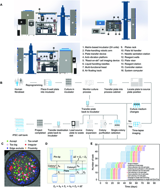
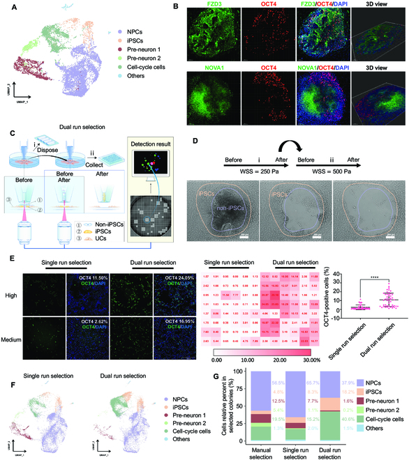
Somatic cell reprogramming generates induced pluripotent stem cells (iPSCs), which serve as a crucial source of seed cells for personalized disease modeling and treatment in regenerative medicine. However, the process of reprogramming often causes substantial lineage manipulations, thereby increasing cellular heterogeneity. As a consequence, the process of harvesting monoclonal iPSCs is labor-intensive and leads to decreased reproducibility. Here, we report the first in-house developed robotic platform that uses a pin-tip-based micro-structure to manipulate radial shear flow for automated monoclonal iPSC colony selection (~1 s) in a non-invasive and label-free manner, which includes tasks for somatic cell reprogramming culturing, medium changes; time-lapse-based high-content imaging; and iPSCs monoclonal colony detection, selection, and expansion. Throughput-wise, this automated robotic system can perform approximately 24 somatic cell reprogramming tasks within 50 days in parallel via a scheduling program. Moreover, thanks to a dual flow-based iPSC selection process, the purity of iPSCs was enhanced, while simultaneously eliminating the need for single-cell subcloning. These iPSCs generated via the dual processing robotic approach demonstrated a purity 3.7 times greater than that of the conventional manual methods. In addition, the automatically produced human iPSCs exhibited typical pluripotent transcriptional profiles, differentiation potential, and karyotypes. In conclusion, this robotic method could offer a promising solution for the automated isolation or purification of lineage-specific cells derived from iPSCs, thereby accelerating the development of personalized medicines.


Conflict of interest statement

Competing interests: The authors declare that they have no competing interests.

Figures

Fig. 1.
Fig. 1.
Utilizing fluid shear stress enables the selection of hiPSCs based on pluripotency-related gene expression patterns. (A) Schematic representation of changes in integrin and cadherin expression patterns during somatic cell reprogramming. (B) The heatmap analysis of ssGSEA results between somatic cells and their derived iPSCs using reference gene sets for integrins, cadherins, and pluripotency, respectively. B: blood, Fib: fibroblast, UC: urine cell. (C) Correlation analysis of pluripotency gene set with integrin gene set or cadherin gene set in somatic cells and their derived iPSCs. (D) Immunofluorescence staining of cadherins (CDH1 and CDH6) in somatic cells (UCs) and their derived iPSCs; scale bar is 20 μm. (E) Schematic of fluid shear stress-induced iPSCs colony dissociation in PPFC. (F) Correlation analysis of iPSC colony dissociation with WSS. (G) Immunofluorescence staining comparison of iPSCs cell–cell connections under static conditions or after dissociation by laminar flow. Scale bar is 10 μm.
Fig. 2.
Fig. 2.
The specificity selection of adhered cells by the PTMS-FLOW. (A) Schematic of pin-tip-based micro-structure (PTMS) for generating localized radial flow and the principle of cell specificity selection. (B) The simulation of the flow field distribution based on the CFD method. The WSS is proportional to uptake flow (Q), whose magnitude is equal to 3μπr0h02Q. The micro-structure allows the pin to aspirate with a constant WSS in the radial direction above the cell culture surface. (C) The experimental principle for PTMS-FLOW-based cell colony selection using the PTMS. (D) Comparison of fluid selected area of NPCs, iPSCs, and UCs after applied PTMS-FLOW. The NPCs illustrate the lowest adhesion force, followed by iPSCs and UCs.
Fig. 3.
Fig. 3.
Robotic system for selecting single iPSCs colony. (A) Mechanical structure and layout of the robotic system for single iPSC colony selection and culturing, shown from different orientations (front, back, and top). The key functional modules of this system include 6 compartments, matrix-based incubator, the plate-handling robotic arm, the cell process cabinet, the reagent station, the controller station, and the system computer, which are labeled with numbers. The precisions of the X, Y, and Z axis of the multi-functional head (the linear motor) are ±0.6 μm/250 mm, ±2.0 μm/250 mm, and ±2.2 μm/50 mm, respectively. (B) The workflow of the iPSC colony selection and culture system. (C) The results of conducting segmentation on a detection map; each resulting segment represents a detected colony. Different colors indicate different maturation periods, which are listed at the top of the map. (D) The principle of an adaptive feedback controlling mechanism is used to ensure the aurate Z-axis distance of the PTMS. (E) Task scheduling. The entire process was managed by programmed scheduling for parallel operation.
Fig. 4.
Fig. 4.
PTMS-FLOW technology enhances iPSC generation by cell fate selection. (A) UMAP embedding showing cell types of the selected colonies through manual selection. NPCs: neuron progenitor cells, iPSCs: induced pluripotent stem cells. (B) Immunofluorescence staining of reprogrammed colonies on Day 15. Top: FZD3 (green), OCT4 (red), and DAPI (blue); bottom: NOVA1 (green), OCT4 (red), and DAPI (blue). The confocal 3-dimensional (3D) datasets were processed with IMARIS software for 3D view; scale bar is 100 μm. (C) Schematic representation of the dual selection mode to obtain colonies. (D) Bright-field images of selected colonies using the dual selection mode. Scale bar is 200 μm. (E) OCT4 staining of selected colonies (left: single selection mode; right: dual selection mode; the OCT4 positive is represented iPSCs); High and Medium represented the fluorescence value of OCT4 staining at the high level and middle level in the corresponding selection mode, respectively. The data on the right side is the quantitative analysis of the fluorescent value of the OCT4 staining. ****P < 0.0001; scale bar is 200 μm. (F) UMAP plot analysis of the cell types from single run selection and dual run selection. (G) Comparison of the proportion of cell types (NPCs, iPSCs, Pre-neuron 1, Pre-neuron 2, Cell-cycle cells, and others) among manual selection mode, single run selection mode, and dual run selection mode.
Fig. 5.
Fig. 5.
Overview of the robotic system for the iPSC colonies selection.

Similar articles

Cited by

References

    1. Yamanaka S. Pluripotent stem cell-based cell therapy-promise and challenges. Cell Stem Cell. 2020;27(4):523–531. - PubMed
    1. Lancaster MA, Knoblich JA. Organogenesis in a dish: Modeling development and disease using organoid technologies. Science. 2014;345(6194):1247125. - PubMed
    1. Lau KX, Mason EA, Kie J, de Souza DP, Kloehn J, Tull D, McConville MJ, Keniry A, Beck T, Blewitt ME, et al. . Unique properties of a subset of human pluripotent stem cells with high capacity for self-renewal. Nat Commun. 2020;11(1):2420. - PMC - PubMed
    1. Tabar V, Studer L. Pluripotent stem cells in regenerative medicine: Challenges and recent progress. Nat Rev Genet. 2014;15(2):82–92. - PMC - PubMed
    1. Deuse T, Hu X, Gravina A, Wang D, Tediashvili G, de C, Thayer WO, Wahl A, Garcia JV, Reichenspurner H, et al. . Hypoimmunogenic derivatives of induced pluripotent stem cells evade immune rejection in fully immunocompetent allogeneic recipients. Nat Biotechnol. 2019;37(3):252–258. - PMC - PubMed