. 2024 Feb 16:15:1352306.
doi: 10.3389/fimmu.2024.1352306. eCollection 2024.

Brucella abortus triggers the differential expression of immunomodulatory lncRNAs in infected murine macrophages

Affiliations

Brucella abortus triggers the differential expression of immunomodulatory lncRNAs in infected murine macrophages

Manuel Flores-Concha et al. Front Immunol. .

Abstract

Introduction: The lncRNAs (long non-coding RNAs) are the most diverse group of non-coding RNAs and are involved in most biological processes including the immune response. While some of them have been recognized for their influence on the regulation of inflammatory activity, little is known in the context of infection by Brucella abortus, a pathogen that presents significant challenges due to its ability to manipulate and evade the host immune system. This study focuses on characterize the expression profile of LincRNA-cox2, Lethe, lincRNA-EPS, Malat1 and Gas5 during infection of macrophages by B. abortus.

Methods: Using public raw RNA-seq datasets we constructed for a lncRNA expression profile in macrophages Brucella-infected. In addition, from public RNA-seq raw datasets of RAW264.7 cells infected with B. abortus we constructed a transcriptomic profile of lncRNAs in order to know the expression of the five immunomodulating lncRNAs studied here at 8 and 24 h post-infection. Finally, we performed in vitro infection assays in RAW264.7 cells and peritoneal macrophages to detect by qPCR changes in the expression of these lncRNAs at first 12 hours post infection, a key stage in the infection cycle where Brucella modulates the immune response to survive.

Results: Our results demonstrate that infection of macrophages with Brucella abortus, induces significant changes in the expression of LincRNA-Cox2, Lethe, LincRNA-EPS, Gas5, and Malat1.

Discussion: The change in the expression profile of these immunomodulatory lncRNAs in response to infection, suggest a potential involvement in the immune evasion strategy employed by Brucella to facilitate its intracellular survival.

Keywords: brucella abortus; gene expression; immunomodulation; lncRNA; macrophages infection.


Conflict of interest statement

The authors declare that the research was conducted in the absence of any commercial or financial relationships that could be construed as a potential conflict of interest.

Figures

Figure 1
Figure 1
Differential expression analysis of lncRNAs in B abortus-infected BMM at 24 h The volcano plots present the results of a differential expression analysis for (A) lncRNA genes and (B) lncRNA transcripts. These were identified by performing bioinformatic analysis on publicly available raw sequencing data (GEO accession: GSE100914) derived from total RNA samples extracted from bone marrow macrophages (BMM) infected with Brucella abortus strain 544. The plots highlight downregulated genes or transcripts in blue and upregulated ones in red, using a log-fold change (LogFC) threshold for color coding. Genes and transcripts that do not exhibit a statistically significant change in expression are depicted in gray based on a significance threshold of 0.05.
Figure 2
Figure 2
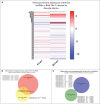
Identification and comparison of the expression of splicing variants of the five evaluated lncRNAs during B abortus-infected RAW264.7 macrophages. The expression of 72 splicing variants of five lncRNAs related to the immune response detected in B abortus-infected RAW264.7 macrophages were analyzed at 8 and 24 h post-infection. (A) Heat Map shows the log2 (fold change) calculated by TPM of each transcript, where blue corresponds to values of downregulated transcripts, and red corresponds to upregulated transcripts, both compared to the uninfected control. The Venn diagram shows the transcript_id, gene_symbol, and log2 (fold change TPM) of splicing variants that are upregulated (B) or downregulated (C).
Figure 3
Figure 3
Interaction networks between immune-related genes and genes correlated with immunomodulatory lncRNAs. Interaction networks of 49 genes related to the lncRNAs analyzed in this study and overexpressed in macrophages infected with B abortus 2308 after 8 h post-infection. The figure shows 73 interactions between genes related to the immune response according to the InnateDB database.
Figure 4
Figure 4
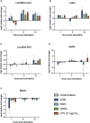
Expression of five lncRNAs related to the regulation of the inflammatory response in B abortus-infected RAW 264.7 macrophages. The transcriptional expression of lincRNA-Cox2 (A), Lethe (B), lincRNA-EPS (C), GAS5 (D), and Malat1 (E) was analyzed using RT-qPCR in RAW 264.7 macrophages infected with B abortus 2308 (2308), B abortus RB51 (RB51), or heat-killed B abortus 2308 (HKBA) at a multiplicity of infection (MOI) of 100:1 for 1, 6, and 12 h LPS was used as the control for the expression of the five lncRNAs. GapdH was used as the reference gene, and significant differences in expression were evaluated compared to the unstimulated control group. Asterisks indicate significant differences in expression compared to the unstimulated control group (*P < 0.05).
Figure 5
Figure 5
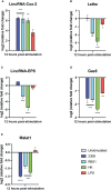
Expression of lncRNAs in peritoneal macrophages infected with B abortus. The expression of (A) lincRNA-Cox2, (B) lincRNA-EPS, (C) Lethe, (D) Gas5, and (E) Malat1 was analyzed by RT-qPCR in peritoneal macrophages treated for 12 hours with S2308, RB51, HKBA (MOI = 100) or LPS. GapdH was used as a reference gene, and significant differences in expression change were compared to the unstimulated control group (*P < 0.05; **P < 0.01; ***P < 0.001; ****P < 0.0001).
Figure 6
Figure 6
Secretion of pro-inflammatory cytokines. The levels of IL-6 (A) and TNF-α (B) secreted by RAW 264.7 macrophages infected with S2308 (2308), RB51, stimulated with HKBA, or stimulated with LPS after 1, 6, and 12 h were quantified by ELISA. Furthermore, the levels of IL-6 (C) and TNF-α (D) secreted in the culture medium of peritoneal macrophages 12 h after being treated with S2308, RB51, HKBA, or LPS were measured by ELISA. The results are expressed as the mean ± standard deviation of three replicates. The asterisks indicate significant differences compared to the unstimulated control (*P < 0.05; **P < 0.01; ***P < 0.001; ****P < 0.0001). The lines under the asterisks with vertical ends indicate differences between the two groups, and the straight lines under the asterisks indicate differences compared to the unstimulated group.

Similar articles

References

    1. Pappas G, Papadimitriou P, Akritidis N, Christou L, Tsianos EV. The new global map of human brucellosis. Lancet Infect Dis (2006) 6:91–9. doi: 10.1016/S1473-3099(06)70382-6 - DOI - PubMed
    1. Wang X, Jiang H. Global prevalence of human brucellosis. Zhonghua Liu Xing Bing Xue Za Zhi (2020) 41:1717–22. doi: 10.3760/cma.j.cn112338-20191022-00751 - DOI - PubMed
    1. Whatmore AM, Foster JT. Emerging diversity and ongoing expansion of the genus Brucella. Infection Genet Evol (2021) 92:104865. doi: 10.1016/j.meegid.2021.104865 - DOI - PubMed
    1. De Figueiredo P, Ficht TA, Rice-Ficht A, Rossetti CA, Adams LG. Pathogenesis and immunobiology of brucellosis: Review of Brucella-host interactions. Am J Pathol (2015) 185:1505–17. doi: 10.1016/j.ajpath.2015.03.003 - DOI - PMC - PubMed
    1. Smith JA. Brucella lipopolysaccharide and pathogenicity: The core of the matter. Virulence (2018) 9:379–82. doi: 10.1080/21505594.2017.1395544 - DOI - PMC - PubMed

Publication types