

Case Reports
doi: 10.1016/j.jdcr.2023.12.016.
eCollection 2024 Mar.
West Nile virus encephalitis presenting with a vesicular dermatitis
Affiliations
- PMID: 38464779
- PMCID: PMC10920127
- DOI: 10.1016/j.jdcr.2023.12.016
Item in Clipboard
Case Reports
West Nile virus encephalitis presenting with a vesicular dermatitis
JAAD Case Rep.
.
No abstract available
Keywords: West Nile virus; encephalitis; flavivirus; immunofluorescence; immunohistochemistry; vesiculobullous eruption.
Conflict of interest statement
None disclosed.
Figures
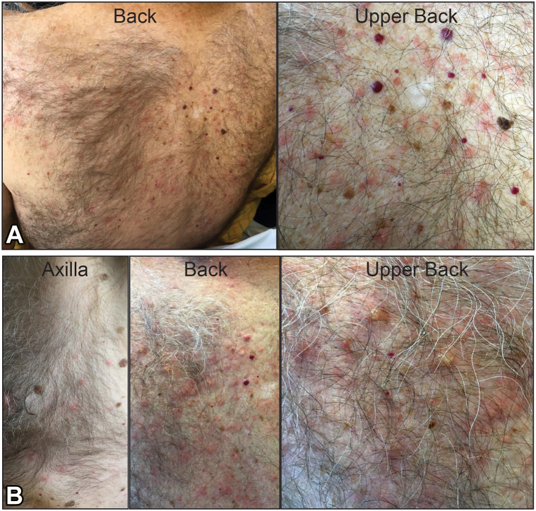
Clinical presentation of (A) initial day 4 and (B) evolving day 6 skin lesions in patient diagnosed with West Nile virus encephalitis. Early lesions were pink macules and papules on the back and chest. Late lesions included erythematous papules and vesicles on the back, chest, and extremities.
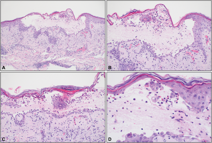
Biopsy revealed an acantholytic intraepidermal vesicle with focal dyskeratosis and a mixed infiltrate in the papillary dermis. (Original magnifications: A, ×100; B, ×200; C, ×200; D, ×400).
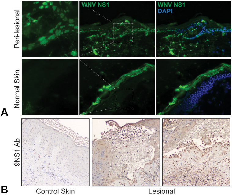
Immunofluorescence and immunohistochemical (IHC) staining for West Nile virus (WNV) NS1 glycoprotein. A, Biopsies were incubated with an antibody against WNV nonstructural glycoprotein NS1 (9NS1) and 4′,6-diamidino-2-phenylindole (DAPI), which highlights nuclei. Biopsy from perilesional skin from a vesicle revealed a intracellular and fibrillar staining pattern from patient with WNV which was absent in normal-appearing skin obtained from a tissue repository. B, IHC staining of WNV NS1 protein revealed robust intracellular staining of keratinocytes in lesional biopsy from patient with WNV which was absent in normal-appearing skin obtained from a tissue repository.
Similar articles
-
[West Nile fever and West Nile encephalitis].Rinsho Shinkeigaku. 2005 Nov;45(11):884-6. Rinsho Shinkeigaku. 2005. PMID: 16447753 Japanese.
-
West Nile Virus and Tick-Borne Encephalitis Virus Are Endemic in Equids in Eastern Austria.Viruses. 2021 Sep 19;13(9):1873. doi: 10.3390/v13091873. Viruses. 2021. PMID: 34578454 Free PMC article.
-
Antibody ratios against NS1 antigens of tick-borne encephalitis and West Nile viruses support differential flavivirus serology in dogs.Transbound Emerg Dis. 2022 Sep;69(5):e2789-e2799. doi: 10.1111/tbed.14630. Epub 2022 Jul 1. Transbound Emerg Dis. 2022. PMID: 35704505
-
[Flavivirus encephalitis: focus on West Nile encephalitis].Rinsho Byori. 2005 Aug;53(8):721-7. Rinsho Byori. 2005. PMID: 16190358 Review. Japanese.
-
Preclinical and clinical development of YFV 17D-based chimeric vaccines against dengue, West Nile and Japanese encephalitis viruses.Vaccine. 2010 Jan 8;28(3):632-49. doi: 10.1016/j.vaccine.2009.09.098. Epub 2009 Oct 4. Vaccine. 2010. PMID: 19808029 Review.
References
-
- McIntosh B., Jupp P., Dos Santos I., Meenehan G. Epidemics of West Nile and Sindbis viruses in South Africa with Culex (Culex) univittatus Theobald as vector. S Afr J Sci. 1976;72:295–300.
-
- Murgue B., Murri S., Triki H., Deubel V., Zeller H.G. West Nile in the Mediterranean basin: 1950-2000. Ann N Y Acad Sci. 2001;951:117–126. - PubMed
-
- Zehender G., Ebranati E., Bernini F., et al. Phylogeography and epidemiological history of West Nile virus genotype 1a in Europe and the Mediterranean basin. Infect Genet Evol. 2011;11(3):646–653. - PubMed
Publication types
Grants and funding
LinkOut - more resources
Full Text Sources