An Optimized P. berghei Liver Stage-HepG2 Infection Model for Simultaneous Quantitative Bioimaging of Host and Parasite Nascent Proteomes
- PMID: 38464937
- PMCID: PMC10917691
- DOI: 10.21769/BioProtoc.4952
An Optimized P. berghei Liver Stage-HepG2 Infection Model for Simultaneous Quantitative Bioimaging of Host and Parasite Nascent Proteomes
Abstract
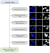
The Plasmodium parasites that cause malaria undergo an obligate, asymptomatic developmental stage in the host liver before initiating the symptomatic blood-stage infection. The parasite liver stage is a key intervention point for antimalarial chemoprophylaxis: successful targeting of liver-stage parasites prevents disease development in individuals and can help to reduce parasite transmission in populations, as the gametocyte forms that transmit infection to mosquitos are exclusively found in the blood stage. Antimalarial drugs that can target multiple parasite stages are thus highly desirable, and one emerging cellular target for such multistage active compounds is the process of protein synthesis or translation. Quantitative study of liver stage translation, and thus mechanistic evaluation of translation inhibitors against liver stage parasites, is not amenable to the methods allowing quantification of asexual blood stage translation, such as radiolabeled amino acid incorporation or lysate-based translation of reporter transcripts. Here, we present a method using o-propargyl puromycin (OPP) labeling of host and parasite nascent proteomes in the P. berghei-HepG2 infection model, followed by automated confocal image acquisition and computational separation of P. berghei vs. H. sapiens nascent proteome signals to allow simultaneous readout of the effects of translation inhibitors on both host and parasite. This protocol details our HepG2 cell culture and infected monolayer handling optimized for microscopy, our OPP labeling workflow, and our approach to automated confocal imaging, image processing, and data analysis. Key features • Uses the o-propargyl puromycin labeling technique developed by Liu et al. to quantitatively analyze protein synthesis in Plasmodium berghei liver-stage parasites in actively translating hepatoma cells. • This quantitative approach should be adaptable for other puromycin-sensitive intracellular pathogens residing in actively translating host cells. • The P. berghei-infected HepG2 recovery and reseeding protocol presented here is of use in applications beyond nascent proteome labeling and quantification.
Keywords: Antimalarial drug discovery; Automated confocal feedback microscopy; HepG2 cell culture; Liver stage; Plasmodium berghei; Protein synthesis; Quantitative bioimaging; Translation.
©Copyright : © 2024 The Authors; This is an open access article under the CC BY-NC license.
Conflict of interest statement
Competing interestsThe authors declare no competing interests.
Figures


Similar articles
-
A high content imaging assay for identification of specific inhibitors of native Plasmodium liver stage protein synthesis.Antimicrob Agents Chemother. 2024 Oct 8;68(10):e0079324. doi: 10.1128/aac.00793-24. Epub 2024 Sep 10. Antimicrob Agents Chemother. 2024. PMID: 39254294 Free PMC article.
-
Single-cell quantitative bioimaging of P. berghei liver stage translation.bioRxiv [Preprint]. 2023 Jul 5:2023.07.05.547872. doi: 10.1101/2023.07.05.547872. bioRxiv. 2023. Update in: mSphere. 2023 Dec 20;8(6):e0054423. doi: 10.1128/msphere.00544-23. PMID: 37461595 Free PMC article. Updated. Preprint.
-
Translation inhibition efficacy does not determine the Plasmodium berghei liver stage antiplasmodial efficacy of protein synthesis inhibitors.bioRxiv [Preprint]. 2023 Dec 7:2023.12.07.570699. doi: 10.1101/2023.12.07.570699. bioRxiv. 2023. Update in: Life Sci Alliance. 2024 Apr 4;7(6):e202302540. doi: 10.26508/lsa.202302540. PMID: 38106175 Free PMC article. Updated. Preprint.
-
Single-cell quantitative bioimaging of P. berghei liver stage translation.mSphere. 2023 Dec 20;8(6):e0054423. doi: 10.1128/msphere.00544-23. Epub 2023 Nov 1. mSphere. 2023. PMID: 37909773 Free PMC article.
-
Advances in omics-based methods to identify novel targets for malaria and other parasitic protozoan infections.Genome Med. 2019 Oct 22;11(1):63. doi: 10.1186/s13073-019-0673-3. Genome Med. 2019. PMID: 31640748 Free PMC article. Review.
Cited by
-
A high content imaging assay for identification of specific inhibitors of native Plasmodium liver stage protein synthesis.bioRxiv [Preprint]. 2024 May 29:2024.05.29.596519. doi: 10.1101/2024.05.29.596519. bioRxiv. 2024. Update in: Antimicrob Agents Chemother. 2024 Oct 8;68(10):e0079324. doi: 10.1128/aac.00793-24. PMID: 38854116 Free PMC article. Updated. Preprint.
-
Differential effects of translation inhibitors on Plasmodium berghei liver stage parasites.Life Sci Alliance. 2024 Apr 4;7(6):e202302540. doi: 10.26508/lsa.202302540. Print 2024 Jun. Life Sci Alliance. 2024. PMID: 38575357 Free PMC article.
-
A high content imaging assay for identification of specific inhibitors of native Plasmodium liver stage protein synthesis.Antimicrob Agents Chemother. 2024 Oct 8;68(10):e0079324. doi: 10.1128/aac.00793-24. Epub 2024 Sep 10. Antimicrob Agents Chemother. 2024. PMID: 39254294 Free PMC article.
References
Grants and funding
LinkOut - more resources
Full Text Sources

