

Apparently X-linked Foveal Hypoplasia of Two Brothers: A Report of a Rare Case
- PMID: 38465154
- PMCID: PMC10924645
- DOI: 10.7759/cureus.53891
Apparently X-linked Foveal Hypoplasia of Two Brothers: A Report of a Rare Case
Abstract
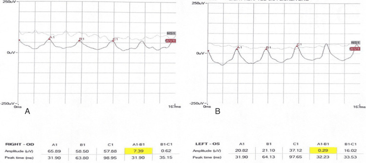
Foveal hypoplasia is a retinal disorder characterized by the anatomic absence of the foveal pit. It might be isolated or associated with poor vision and several conditions such as albinism, aniridia, microphthalmos, congenital nystagmus, or other diseases. Genetic and non-genetic causes can play a role in foveal pit development. However, the exact mechanism that causes foveal pit absence has not been determined. This study reports a five-year-old boy who presented to the eye clinic with bilateral poor vision since birth. Optical coherence tomography (OCT) was performed and confirmed the absence of the foveal pit in both eyes. Diagnosis of foveal hypoplasia was made. The parents reported a positive family history of similar conditions, specifically, a paternal grandfather, a male paternal cousin, and a brother. To the best of our knowledge, this is the first reported case of foveal hypoplasia, with a positive family history in the male gender specifically. Thus, inheritance is presumed to be X-linked recessive. We acknowledge that further investigation by genetic testing would offer further insight into this case.
Keywords: case report; foveal hypoplasia; nystagmus; optical coherence tomography (oct); visual acuity.
Copyright © 2024, Alarfaj et al.
Conflict of interest statement
The authors have declared that no competing interests exist.
Figures


Similar articles
-
Nystagmus associated with macular dysplasia.Strabismus. 2020 Mar;28(1):17-19. doi: 10.1080/09273972.2019.1668028. Epub 2019 Sep 30. Strabismus. 2020. PMID: 31566469
-
Case Report: Optical Coherence Tomography Angiography of Idiopathic Foveal Hypoplasia and Its Correlation With Visual Acuity.Optom Vis Sci. 2020 Feb;97(2):110-120. doi: 10.1097/OPX.0000000000001471. Optom Vis Sci. 2020. PMID: 32011584
-
Spectral-domain optical coherence tomography foveal morphology as a prognostic factor for vision performance in congenital aniridia.Eur J Ophthalmol. 2020 Jan;30(1):58-65. doi: 10.1177/1120672118818352. Epub 2018 Dec 17. Eur J Ophthalmol. 2020. PMID: 30556423
-
Foveal hypoplasia and optical coherence tomographic imaging.Taiwan J Ophthalmol. 2018 Oct-Dec;8(4):181-188. doi: 10.4103/tjo.tjo_101_18. Taiwan J Ophthalmol. 2018. PMID: 30637189 Free PMC article. Review.
-
Autosomal dominant foveal hypoplasia without visible macular abnormalities and PAX6 mutations.Jpn J Ophthalmol. 2020 Nov;64(6):635-641. doi: 10.1007/s10384-020-00766-9. Epub 2020 Aug 28. Jpn J Ophthalmol. 2020. PMID: 32857266 Review.
References
-
- Variable clinical profile of fovea plana in normal children. Villegas VM, Schwartz SG, Hamet TD, McKeown CA, Capó H, Flynn HW Jr. Ophthalmic Surg Lasers Imaging Retina. 2018;49:251–257. - PubMed
Publication types
LinkOut - more resources
Full Text Sources