

Inverted apicobasal polarity in health and disease
- PMID: 38465512
- PMCID: PMC10984280
- DOI: 10.1242/jcs.261659
Inverted apicobasal polarity in health and disease
Abstract
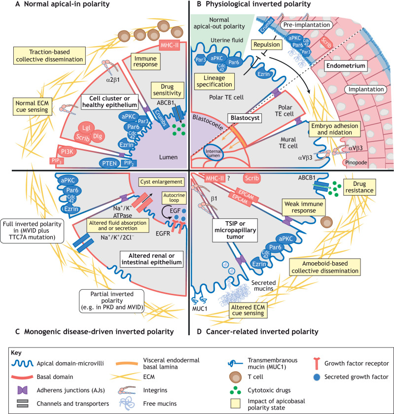
Apicobasal epithelial polarity controls the functional properties of most organs. Thus, there has been extensive research on the molecular intricacies governing the establishment and maintenance of cell polarity. Whereas loss of apicobasal polarity is a well-documented phenomenon associated with multiple diseases, less is known regarding another type of apicobasal polarity alteration - the inversion of polarity. In this Review, we provide a unifying definition of inverted polarity and discuss multiple scenarios in mammalian systems and human health and disease in which apical and basolateral membrane domains are interchanged. This includes mammalian embryo implantation, monogenic diseases and dissemination of cancer cell clusters. For each example, the functional consequences of polarity inversion are assessed, revealing shared outcomes, including modifications in immune surveillance, altered drug sensitivity and changes in adhesions to neighboring cells. Finally, we highlight the molecular alterations associated with inverted apicobasal polarity and provide a molecular framework to connect these changes with the core cell polarity machinery and to explain roles of polarity inversion in health and disease. Based on the current state of the field, failure to respond to extracellular matrix (ECM) cues, increased cellular contractility and membrane trafficking defects are likely to account for most cases of inverted apicobasal polarity.
Keywords: Apicobasal polarity; Embryo implantation; Extracellular matrix sensing; Membrane trafficking; Micropapillary cancer; Monogenic diseases.
© 2024. Published by The Company of Biologists Ltd.
Conflict of interest statement
Competing interests F.J. is the CEO of a Gustave Roussy spin-off (Orakl).
Figures

Similar articles
-
Apicobasal Surfaceome Architecture Encodes for Polarized Epithelial Functionality and Depends on Tumor Suppressor PTEN.Int J Mol Sci. 2022 Dec 19;23(24):16193. doi: 10.3390/ijms232416193. Int J Mol Sci. 2022. PMID: 36555834 Free PMC article.
-
Tuning apicobasal polarity and junctional recycling in the hemogenic endothelium orchestrates the morphodynamic complexity of emerging pre-hematopoietic stem cells.Elife. 2024 May 29;12:RP91429. doi: 10.7554/eLife.91429. Elife. 2024. PMID: 38809590 Free PMC article.
-
Apico-basal polarity in polycystic kidney disease epithelia.Biochim Biophys Acta. 2011 Oct;1812(10):1239-48. doi: 10.1016/j.bbadis.2011.05.008. Epub 2011 Jun 1. Biochim Biophys Acta. 2011. PMID: 21658447 Review.
-
Apicobasal polarity in the kidney.Exp Cell Res. 2012 May 15;318(9):1033-9. doi: 10.1016/j.yexcr.2012.02.028. Epub 2012 Mar 6. Exp Cell Res. 2012. PMID: 22421511 Free PMC article. Review.
-
New insights into the organization and regulation of the apical polarity network in mammalian epithelial cells.FEBS J. 2021 Dec;288(24):7073-7095. doi: 10.1111/febs.15710. Epub 2021 Feb 1. FEBS J. 2021. PMID: 33448150 Review.
Cited by
-
Differential Effects of Confinement on the Dynamics of Normal and Tumor-Derived Pancreatic Ductal Organoids.ACS Appl Bio Mater. 2024 Dec 16;7(12):8489-8502. doi: 10.1021/acsabm.4c01301. Epub 2024 Nov 22. ACS Appl Bio Mater. 2024. PMID: 39576883 Free PMC article.
-
A Patient-Derived 3D Cyst Model of Polycystic Kidney Disease That Mimics Disease Development and Responds to Repurposing Candidates.Clin Transl Sci. 2025 Apr;18(4):e70214. doi: 10.1111/cts.70214. Clin Transl Sci. 2025. PMID: 40235151 Free PMC article.
-
Changes in Epithelial Cell Polarity and Adhesion Guide Human Endometrial Receptivity: How In Vitro Systems Help to Untangle Mechanistic Details.Biomolecules. 2025 Jul 22;15(8):1057. doi: 10.3390/biom15081057. Biomolecules. 2025. PMID: 40867502 Free PMC article. Review.
References
-
- Al-Obaidy, K. I., Eble, J. N., Cheng, L., Williamson, S. R., Sakr, W. A., Gupta, N., Idrees, M. T. and Grignon, D. J. (2019). Papillary renal neoplasm with reverse polarity: a morphologic, immunohistochemical, and molecular study. Am. J. Surg. Pathol. 43, 1099-1111. 10.1097/PAS.0000000000001288 - DOI - PubMed
Publication types
MeSH terms
Grants and funding
LinkOut - more resources
Full Text Sources
