

Relationship of Perivascular Space Markers With Incident Dementia in Cerebral Small Vessel Disease
- PMID: 38465597
- PMCID: PMC10962441
- DOI: 10.1161/STROKEAHA.123.045857
Relationship of Perivascular Space Markers With Incident Dementia in Cerebral Small Vessel Disease
Abstract
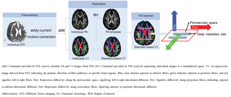
Background: Recent studies, using diffusion tensor image analysis along the perivascular space (DTI-ALPS), suggest impaired perivascular space (PVS) function in cerebral small vessel disease, but they were cross-sectional, making inferences on causality difficult. We determined associations between impaired PVS, measured using DTI-ALPS and PVS volume, and cognition and incident dementia.
Methods: In patients with lacunar stroke and confluent white matter hyperintensities, without dementia at baseline, recruited prospectively in a single center, magnetic resonance imaging was performed annually for 3 years, and cognitive assessments, including global, memory, executive function, and processing speed, were performed annually for 5 years. We determined associations between DTI-ALPS and PVS volume with cerebral small vessel disease imaging markers (white matter hyperintensity volume, lacunes, and microbleeds) at baseline and with changes in imaging markers. We determined whether DTI-ALPS and PVS volume at baseline and change over 3 years predicted incident dementia. Analyses were controlled for conventional diffusion tensor image metrics using 2 markers (median mean diffusivity [MD] and peak width of skeletonized MD) and adjusted for age, sex, and vascular risk factors.
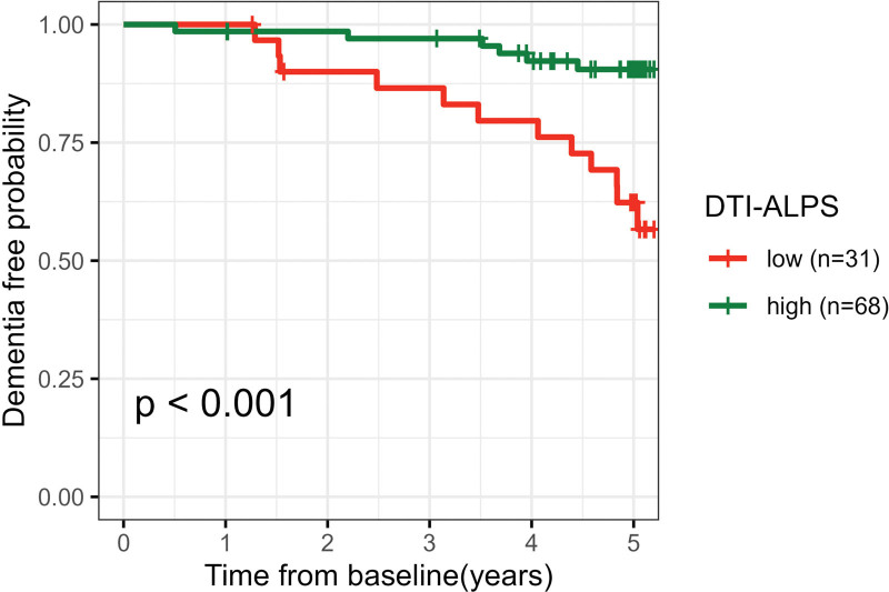
Results: A total of 120 patients, mean age 70.0 years and 65.0% male, were included. DTI-ALPS declined over 3 years, while no change in PVS volume was found. Neither DTI-ALPS nor PVS volume was associated with cerebral small vessel disease imaging marker progression. Baseline DTI-ALPS was associated with changes in global cognition (β=0.142, P=0.032), executive function (β=0.287, P=0.027), and long-term memory (β=0.228, P=0.027). Higher DTI-ALPS at baseline predicted a lower risk of dementia (hazard ratio, 0.328 [0.183-0.588]; P<0.001), and this remained significant after including median MD as a covariate (hazard ratio, 0.290 [0.139-0.602]; P<0.001). Change in DTI-ALPS predicted dementia conversion (hazard ratio, 0.630 [0.428-0.964]; P=0.048), but when peak width of skeletonized MD and median MD were entered as covariates, the association was not significant. There was no association between baseline PVS volume, or PVS change over 3 years, and conversion to dementia.
Conclusions: DTI-ALPS predicts future dementia risk in patients with lacunar strokes and confluent white matter hyperintensities. However, the weakening of the association between change in DTI-ALPS and incident dementia after controlling for peak width of skeletonized MD and median MD suggests part of the signal may represent conventional diffusion tensor image metrics. PVS volume is not a predictor of future dementia risk.
Keywords: cerebral small vessel disease; cognitive dysfunction; dementia; white matter.
Conflict of interest statement
Figures
References
-
- Wardlaw JM, Benveniste H, Nedergaard M, Zlokovic BV, Mestre H, Lee H, Doubal FN, Brown R, Ramirez J, MacIntosh BJ, et al. ; Colleagues From the Fondation Leducq Transatlantic Network of Excellence on the Role of the Perivascular Space in Cerebral Small Vessel Disease. Perivascular spaces in the brain: anatomy, physiology and pathology. Nat Rev Neurol. 2020;16:137–153. doi: 10.1038/s41582-020-0312-z - PubMed
Publication types
MeSH terms
Grants and funding
LinkOut - more resources
Full Text Sources
Medical
Research Materials
