. 2024 Mar 11;19(3):e0295381.
doi: 10.1371/journal.pone.0295381. eCollection 2024.

Chromolaena odorata layered-nitrile rubber polymer transdermal patch enhanced wound healing in vivo

Affiliations

Chromolaena odorata layered-nitrile rubber polymer transdermal patch enhanced wound healing in vivo

Mazlyzam Abdul Latif et al. PLoS One. .

Abstract

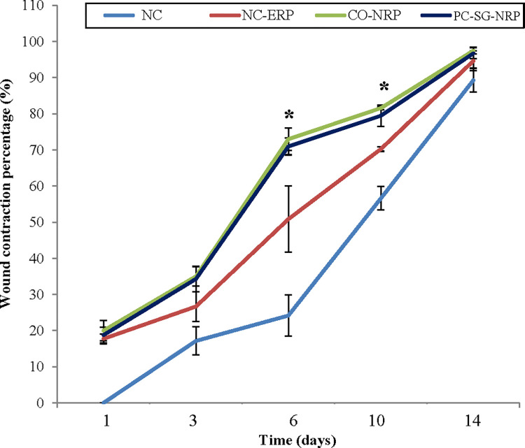
The objective is to investigate the healing efficacy of a Chromolaena odorata layered-nitrile rubber transdermal patch on excision wound healing in rats. Wounds were induced in Sprague-Dawley rats and were later treated as follows: wound A, the negative control, received no treatment (NC); wound B, the negative control with an empty nitrile rubber patch (NC-ERP); wound C, treated with a C. odorata layered-nitrile rubber patch (CO-NRP); and wound D, the positive control with Solcoseryl gel with a nitrile rubber patch (PC-SG-NRP). After 1, 3, 6, 10, and 14 days, the rats were sacrificed and analyzed for wound contraction, protein content, hexosamine, and uronic acid levels. Macroscopic observation showed enhanced wound healing in wounds treated with CO-NRP with a wound contraction percentage significantly higher (p<0.05) on days 6 and 10 compared to those treated with NC-ERP. Similarly, protein, hexosamine, and uronic acid contents were also significantly higher (p<0.05) in CO-NRP-treated wounds when compared with wounds treated with NC-ERP. Histological findings showed denser collagen deposition and faster granulation tissue formation in wounds treated with CO-NRP. From the results obtained, it is concluded that the C. odorata layered-nitrile rubber transdermal patch was effective in healing skin wounds.


Conflict of interest statement

The authors have declared that no competing interests exist.

Figures

Fig 1
Fig 1. Six excisional wounds were induced on the dorsum of rats on day 0.
(I) Wound A, the control, received no treatment (NC). Wound B was the control treated with an empty nitrile rubber patch (NC-ERP). Wound C was treated with a C. odorata layered-nitrile rubber patch (CO-NRP). Wound D, which acted as the positive control, was treated with Solcoseryl gel and a nitrile rubber patch (PC-SG-NRP).
Fig 2
Fig 2. Excisional wound closures in rats as a function of wound treatment and time.
Fig 3
Fig 3. Effect of transdermal patches on the level of wound contraction percentage in an excision wound model.
n = 6 rats in each group; values are mean ± SEM, *Significant at p < 0.05 when wound treated with CO-NRP was compared with wound treated with empty nitrile rubber patch (NC-ERP).
Fig 4
Fig 4. Effect of different treatments on the protein content of excision wounds as a function of time.
n = 6 in each group; values are mean ± SEM, *Significant at p < 0.05 when CO-NRP treated wound was compared with NC-ERP treated wound.
Fig 5
Fig 5. Effect of different treatments on the hexosamines content of excision wounds as a function of time.
n = 6 rats in each group; values are mean ± SEM, *Significant at p < 0.05 when wound C was compared to wound B.
Fig 6
Fig 6. Effect of different treatments on the uronic acid content of excision wounds as a function of time.
n = 6 rats in each group; values are mean ± SEM, *Significant at p < 0.05 when wound C was compared with wound B.
Fig 7
Fig 7. Images of skin tissue sections stained with haematoxylin and eosin show histological changes during the wound healing in untreated and treated wounds as a function of time.
Magnification:100x. Note: Ad = adipose tissue, Bc = blood clot, Ca = capillaries, E = epithelium, Ex = exudates, F = hair follicle, Fi = fibroblasts, G = granulation tissue, I = acute inflammatory cells, M = muscles layers, N = necrotic slough, RBC = red blood cells.
Fig 8
Fig 8. Images of skin tissue sections stained with Masson’s Trichrome showing histopathological changes during the wound healing process in untreated and treated wounds as a function of time.
Magnification: 100x. Note: Ad = adipose tissue, Bc = blood clot, Ca = capillaries, E = epithelium, Ex = exudates, F = hair follicle, Fi = fibroblasts, Ko = collagen, Ko I = type-I collagen, Ko III = type-III collagen, I = acute inflammatory cells, M = muscles layers, N = necrotic slough.

References

    1. desJardins-Park HE, Gurtner GC, Wan DC, Longaker MT. From Chronic Wounds to Scarring: The Growing Health Care Burden of Under- and Over-Healing Wounds. Adv Wound Care (New Rochelle). 2022. Sep;11(9):496–510. doi: 10.1089/wound.2021.0039 Epub 2021 Nov 15. ; PMCID: PMC9634983. - DOI - PMC - PubMed
    1. Woo SH, Choi JH, Mo YJ, Lee YI, Jeon WB, Lee YS. Engineered elastin-like polypeptide improves the efficiency of adipose-derived stem cell-mediated cutaneous wound healing in type II diabetes mellitus. Heliyon. 2023. Sep 15;9(9):e20201. doi: 10.1016/j.heliyon.2023.e20201 ; PMCID: PMC10559957. - DOI - PMC - PubMed
    1. Takeo M, Lee W, Ito M. Wound healing and skin regeneration. Cold Spring Harbor Perspectives in Medicine. 2015;5(1):a023267. Epub 20150105. doi: 10.1101/cshperspect.a023267 ; PubMed Central PMCID: PMC4292081. - DOI - PMC - PubMed
    1. Gonzalez AC, Costa TF, Andrade ZA, Medrado AR. Wound healing—A literature review. Anais Brasileiros de Dermatologia. 2016;91(5):614–20. doi: 10.1590/abd1806-4841.20164741 ; PubMed Central PMCID: PMC5087220. - DOI - PMC - PubMed
    1. Rodrigues M, Kosaric N, Bonham CA, Gurtner GC. Wound Healing: A Cellular Perspective. Physiological Reviews. 2019;99(1):665–706. doi: 10.1152/physrev.00067.2017 ; PubMed Central PMCID: PMC6442927. - DOI - PMC - PubMed