. 2024 Mar 19;121(12):e2316491121.
doi: 10.1073/pnas.2316491121. Epub 2024 Mar 11.

Structure of RADX and mechanism for regulation of RAD51 nucleofilaments

Affiliations

Structure of RADX and mechanism for regulation of RAD51 nucleofilaments

Swati Balakrishnan et al. Proc Natl Acad Sci U S A. .

Abstract

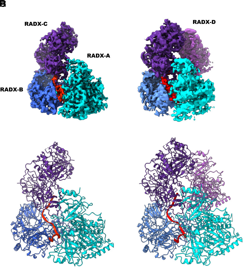
Replication fork reversal is a fundamental process required for resolution of encounters with DNA damage. A key step in the stabilization and eventual resolution of reversed forks is formation of RAD51 nucleoprotein filaments on exposed single strand DNA (ssDNA). To avoid genome instability, RAD51 filaments are tightly controlled by a variety of positive and negative regulators. RADX (RPA-related RAD51-antagonist on the X chromosome) is a recently discovered negative regulator that binds tightly to ssDNA, directly interacts with RAD51, and regulates replication fork reversal and stabilization in a context-dependent manner. Here, we present a structure-based investigation of RADX's mechanism of action. Mass photometry experiments showed that RADX forms multiple oligomeric states in a concentration-dependent manner, with a predominance of trimers in the presence of ssDNA. The structure of RADX, which has no structurally characterized orthologs, was determined ab initio by cryo-electron microscopy (cryo-EM) from maps in the 2 to 4 Å range. The structure reveals the molecular basis for RADX oligomerization and the coupled multi-valent binding of ssDNA binding. The interaction of RADX with RAD51 filaments was imaged by negative stain EM, which showed a RADX oligomer at the end of filaments. Based on these results, we propose a model in which RADX functions by capping and restricting the end of RAD51 filaments.

Keywords: DNA replication; RAD51; cryo-EM; replication fork.


Conflict of interest statement

Competing interests statement:The authors declare no competing interest.

Figures

Fig. 1.
Fig. 1.
RADX oligomerization is stabilized and preferentially forms trimers upon binding ssDNA. (A) RADX is primarily monomeric at 50 nM, but dimerizes at 100 nM. (B) Addition of ssDNA to monomeric RADX leads to a significant increase in the population of trimers, from 4 to 33%. (C and D) Addition of different lengths of ssDNA leads to varying distribution of oligomers. (E) DNA binding affinity of RADX determined by fluorescence polarization anisotropy show there is no dependence on the length of the substrate. (F) Tryptophan quenching assay measuring the DNA footprint of RADX shows a footprint length of 19 to 27 nucleotides.
Fig. 2.
Fig. 2.
High resolution structure of RADX. (A and B) EM map and ribbon representation of the structure of the RADX trimer bound to ssDNA (red) with the three protomers in cyan, blue and purple respectively. (C and D) EM map and ribbon representation of the structure of the RADX tetramer bound to ssDNA (red) with the four protomers in cyan, blue, purple and violet respectively.
Fig. 3.
Fig. 3.
RADX oligomerization is stabilized by multiple inter-domain interfaces. The OB1–OB4 interface (in gold) between two RADX protomers A and B (teal and blue, respectively). RADX oligomerizes primarily via hydrogen bond and salt-bridge interactions between domains 1 and 4. The Left Inset shows the residues involved in hydrogen bonding colored according to the protomer of origin, with the hydrogen bonds shown as red dashed lines. The Right Inset shows the three sites of RADX oligomerization mutations (residues in red) in the central beta sheet of OB4. Mutation of these residues are likely to perturb the beta sheet and the entire D4 domain, which could contribute to effects observed in functional assays.
Fig. 4.
Fig. 4.
RADX binding of ssDNA is coupled to oligomerization. The Top panel shows the ribbon representation (Left) of a RADX protomer (blue) with ssDNA bound (red), with the corresponding map of the surface electrostatic charge showing the positively charged DNA binding surface in blue (Right). The Top Inset shows the consensus residues involved in hydrogen bonding or pi-stacking interactions (blue) with the DNA (red). The Lower Inset shows the sites of the Ob2m mutations (green and orange). Among the mutations, only the two residues with orange labels (W279, K305) have any overlap with the DNA binding site.
Fig. 5.
Fig. 5.
RADX variants show coupling of oligomerization and DNA binding. (AD) RADX-OLM and RADX-DBM do not oligomerize in the presence of ssDNA. (EG) DNA binding affinity of RADX-OLM is significantly lower than wt RADX, while RADX-DBM shows no detectable binding to ssDNA. (H) Measurement of tryptophan quenching of RADX-OLM does not show stoichiometric binding, as compared to wt RADX.
Fig. 6.
Fig. 6.
RADX modulation of RAD51 filaments correlates with end-binding. (A) (Left) Insets from negative stain EM micrographs showing anti-RADX Ab bound gold nanoparticles localized at the ends of RAD51 filaments. (Right) 2D class averages containing RADX crosslinked to RAD51 filaments, which reveal populations of free RADX and RADX bound to RAD51 filaments. (B) 3D reconstructions of free RADX and RADX bound to RAD51 filaments. The extra volume assigned to RADX is highlighted by the red circle. (C) The residues mutated in the RAD51 binding-deficient QVPK mutant displayed on the structure of RADX. These residues form a surface-exposed patch, consistent with the proposal that these are in the RAD51 binding site. (D) Model of a RADX trimer bound to a RAD51 filament generated via manual docking. The RADX is colored green, the RAD51 filament (8GYK) purple, and the ssDNA red.
Fig. 7.
Fig. 7.
Model for the mechanism of RADX action. (A) Upon replication fork stalling, single stranded DNA is exposed, (B) which is then bound by RPA to protect the ssDNA from damaging agents. (C) RPA is replaced on ssDNA with RAD51 by the action of mediator protein BRCA2. RAD51 binds cooperatively to form filaments on ssDNA. (D) RADX is recruited to ssDNA where it binds the ends of the growing RAD51 filament. (E) RADX promotes filament disassembly by either i) blocking expansion at the end of the filament as RAD51 hydrolyses ATP and detaches from ssDNA or ii) both blocking filament expansion and accelerating the ATP hydrolysis rate of RAD51.

Update of

References

    1. Berti M., Cortez D., Lopes M., The plasticity of DNA replication forks in response to clinically relevant genotoxic stress. Nat. Rev. Mol. Cell Biol. 21, 633–651 (2020). - PubMed
    1. Cortez D., Replication-coupled DNA repair. Mol. Cell 74, 866–876 (2019). - PMC - PubMed
    1. Zellweger R., et al. , Rad51-mediated replication fork reversal is a global response to genotoxic treatments in human cells. J. Cell Biol. 208, 563–579 (2015). - PMC - PubMed
    1. Hashimoto Y., Chaudhuri A. R., Lopes M., Costanzo V., Rad51 protects nascent DNA from Mre11-dependent degradation and promotes continuous DNA synthesis. Nat. Struct. Mol. Biol. 17, 1305–1311 (2010). - PMC - PubMed
    1. Schlacher K., et al. , Double-strand break repair-independent role for BRCA2 in blocking stalled replication fork degradation by MRE11. Cell 145, 529–542 (2011). - PMC - PubMed

MeSH terms

LinkOut - more resources