. 2024 Jun:376:114749.
doi: 10.1016/j.expneurol.2024.114749. Epub 2024 Mar 11.

Characterization of the intrahippocampal kainic acid model in female mice with a special focus on seizure suppression by antiseizure medications

Affiliations

Characterization of the intrahippocampal kainic acid model in female mice with a special focus on seizure suppression by antiseizure medications

Melanie Widmann et al. Exp Neurol. 2024 Jun.

Abstract

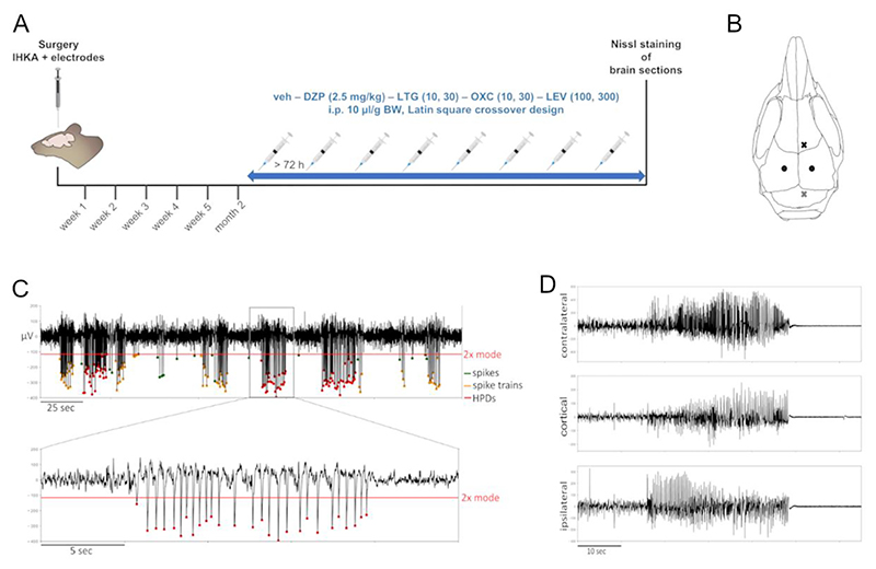
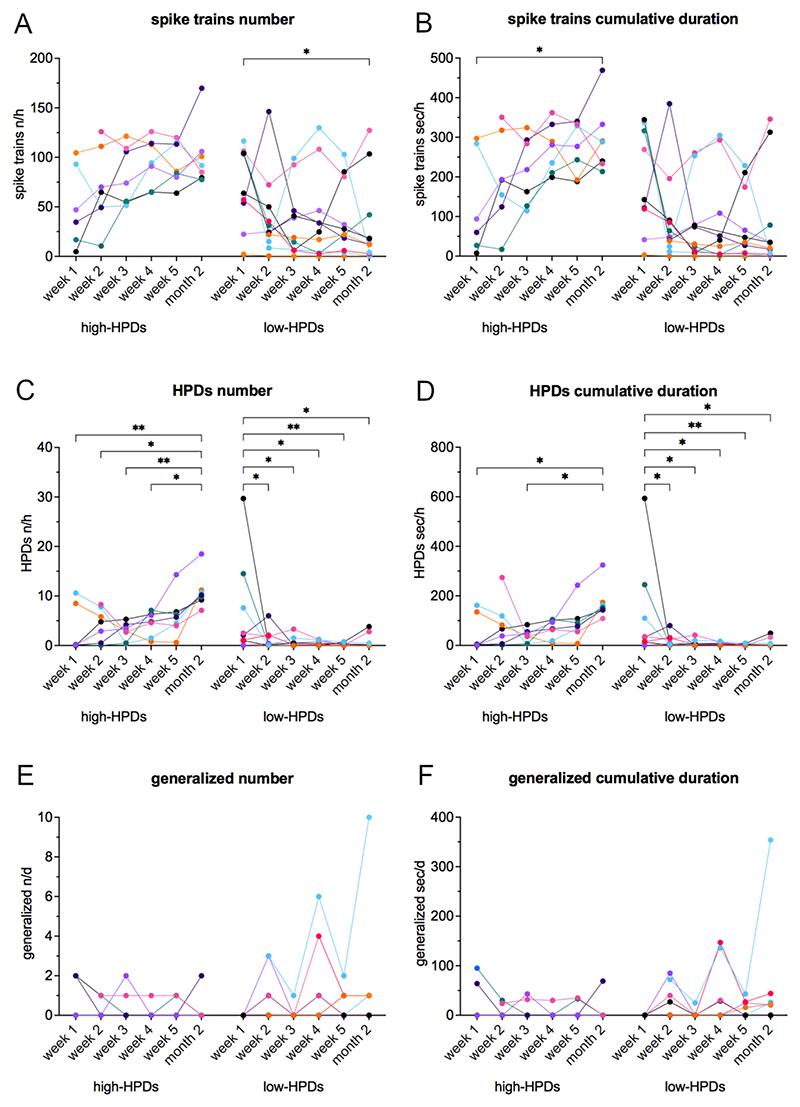
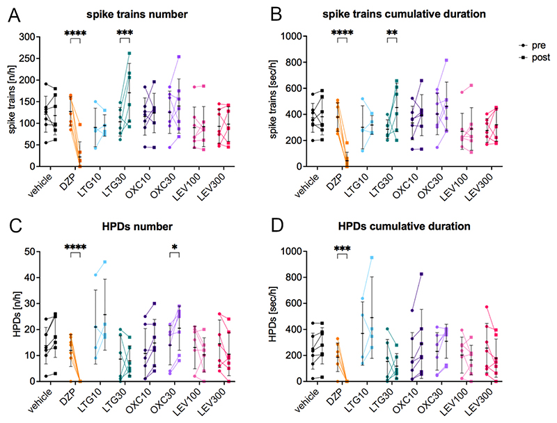
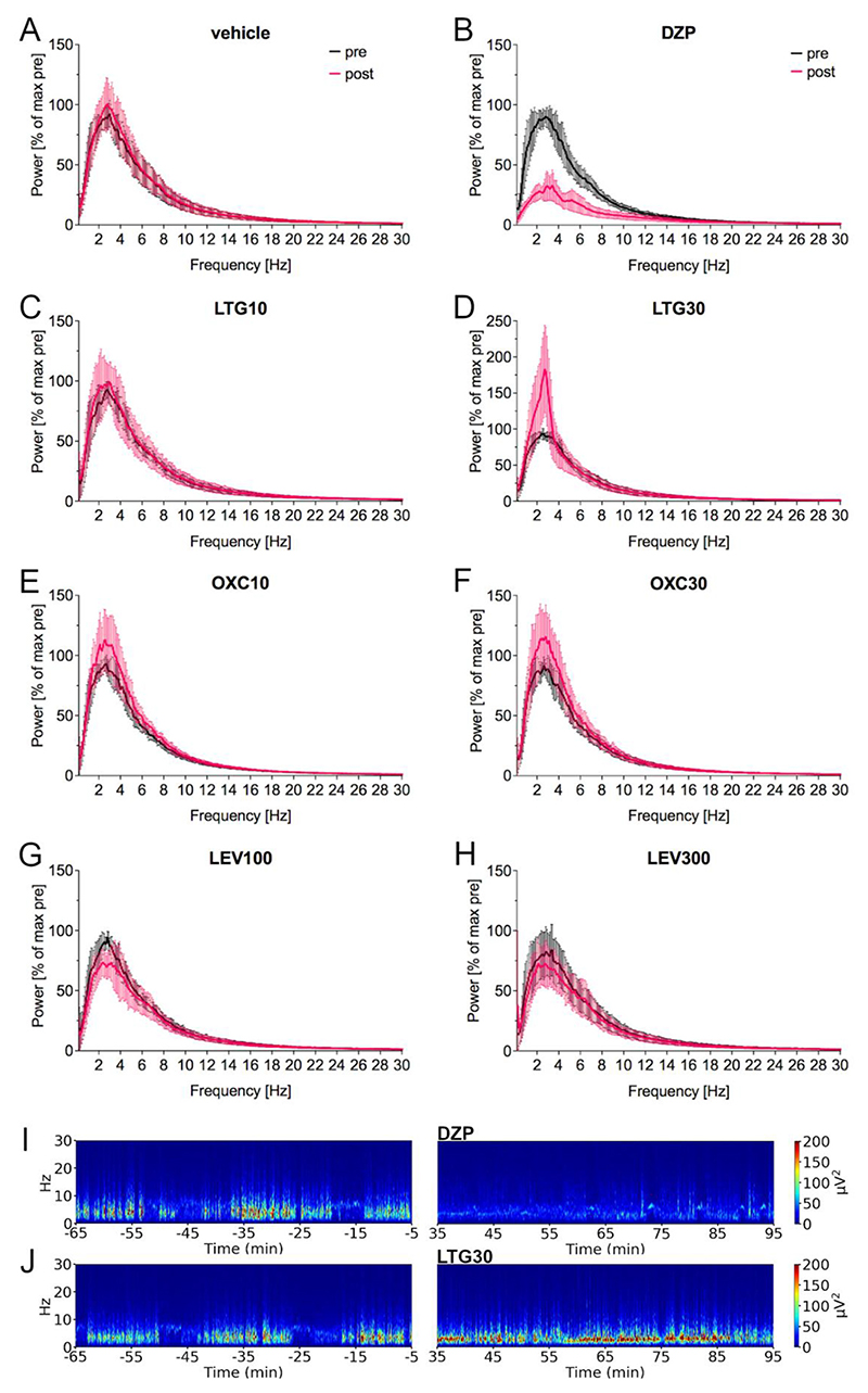
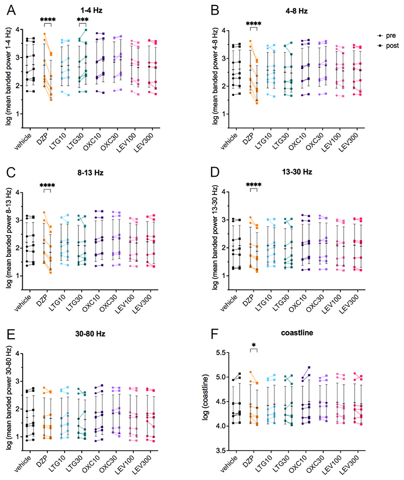
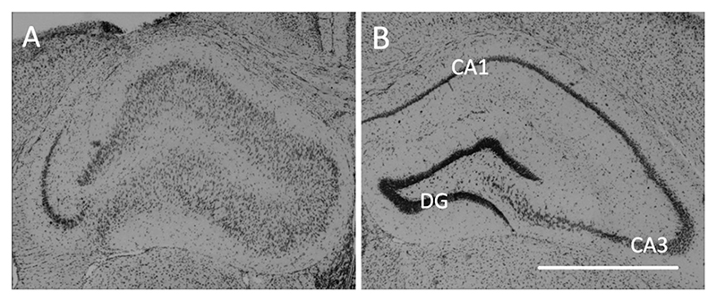
Despite special challenges in the medical treatment of women with epilepsy, in particular preclinical animal studies were focused on males for decades and females have only recently moved into the focus of scientific interest. The intrahippocampal kainic acid (IHKA) mouse model of temporal lobe epilepsy (TLE) is one of the most studied models in males reproducing electroencephalographic (EEG) and histopathological features of human TLE. Hippocampal paroxysmal discharges (HPDs) were described as drug resistant focal seizures in males. Here, we investigated the IHKA model in female mice, in particular drug-resistance of HPDs and the influence of antiseizure medications (ASMs) on the power spectrum. After injecting kainic acid (KA) unilaterally into the hippocampus of female mice, we monitored the development of epileptiform activity by local field potential (LFP) recordings. Subsequently, we evaluated the effect of the commonly prescribed ASMs lamotrigine (LTG), oxcarbazepine (OXC) and levetiracetam (LEV), as well as the benzodiazepine diazepam (DZP) with a focus on HPDs and power spectral analysis and assessed neuropathological alterations of the hippocampus. In the IHKA model, female mice replicated key features of human TLE as previously described in males. Importantly, HPDs in female mice did not respond to commonly prescribed ASMs in line with the drug-resistance in males, thus representing a suitable model of drug-resistant seizures. Intriguingly, we observed an increased occurrence of generalized seizures after LTG. Power spectral analysis revealed a pronounced increase in the delta frequency range after the higher dose of 30 mg/kg LTG. DZP abolished HPDs and caused a marked reduction over a wide frequency range (delta, theta, and alpha) of the power spectrum. By characterizing the IHKA model of TLE in female mice we address an important gap in basic research. Considering the special challenges complicating the therapeutic management of epilepsy in women, inclusion of females in preclinical studies is imperative. A well-characterized female model is a prerequisite for the development of novel therapeutic strategies tailored to sex-specific needs and for studies on the effect of epilepsy and ASMs during pregnancy.

Keywords: Drug-resistant seizures; Hippocampal paroxysmal discharges; Lamotrigine; Levetiracetam; Oxcarbazepine; Temporal lobe epilepsy.


Conflict of interest statement

Declaration of competing interest C. Schwarzer is co-founder of EpiBlok Therapeutics GmbH. None of the other authors has any conflict of interest to disclose. We confirm that we have read the Journal's position on issues involved in ethical publication and affirm that this report is consistent with those guidelines.

Figures

Fig. 1
Fig. 1. Experimental timeline, schematic of the electrode implantation sites and representative LFP traces.
(A) Experimental timeline for monitoring the development of epileptiform activity following IHKA in female mice. Females with established chronic epilepsy (>50 s/h ipsilateral HPDs) were subsequently tested with DZP (2.5 mg/kg), LTG (10 and 30 mg/kg), OXC (10 and 30 mg/kg) and LEV (100 and 300 mg/kg) compared to vehicle (10% DMSO and 90% saline with 3% Tween 80) i.p. in a volume of 10 μl/g. Treatment schedules were designed according to a Latin square crossover design with 72 h minimum resting period between injections. Afterwards, Nissl-stained brain sections were analyzed for morphological alterations. IHKA, intrahippocampal kainic acid; veh, vehicle; DZP, diazepam; LTG, lamotrigine; OXC, oxcarbazepine; LEV, levetiracetam; i.p., intraperitoneal; BW, body weight. (B) Schematic figure showing the electrode implantation sites. The black dots symbolize the 2 depth electrodes targeting the ipsilateral and the contralateral hippocampus (AP -1.8, ML ±1.3, DV -1.8). The black cross represents the single surface electrode implanted above the motor cortex, the grey cross the triple electrode for grounding and references positioned above the cerebellum. (C) Representative LFP trace of the ipsilateral hippocampus showing single spikes, spike trains and HPDs. Epileptiform spikes are characterized by an amplitude 2× mode (green dots represent single spikes). Spike trains (marked with orange dots) are series of at least 3 epileptiform spikes lasting 1–10 s with ≥1.33 Hz. Trains of epileptiform spikes ≥10 s are counted as HPDs (red dots). In this exemplary trace the spike polarity is negative, a minority of animals, however, present with positive spikes. Red line = baseline for spike threshold detection set to amplitude minimum of 2× mode. (D) LFP traces of a typical generalized seizure with approximately 30 s of high-amplitude, high-frequency discharges simultaneously both in the ipsi- and contralateral hippocampus as well as in the cortical electrode followed by a pronounced post-ictal depression of LFP activity.
Fig. 2
Fig. 2. Development of epileptiform activity in the ipsilateral hippocampus in female mice after IHKA.
(A-F) Separated into high-HPDs group (>50 s/h HPDs at 2 months, n = 7) and low-HPDs group (<50 s/h HPDs at 2 months, n = 11), (A) number [n/h] and (B) cumulative duration [s/h] of spike trains, (C) number [n/h] and (D) cumulative duration [s/h] of HPDs, (E) number [n/d] and (F) cumulative duration [s/d] of generalized seizures were assessed during weekly 24 h recordings for the first 5 weeks and at 2 months. Females of the high-HPDs group showed a significant increase of HPDs and a tendency to increase spike trains, while HPDs significantly decreased and spike trains tended to decrease in the low-HPDs group. Regarding generalized seizures, no significant differences were detected. Note one animal of the low-HPDs group with strikingly higher generalized seizure activity increasing up to 10 generalized seizures at the 2 months recording time point. Curves represent individual animals. Two-way mixed-effects model for repeated measures followed by Tukey’s multiple comparisons test (significance levels shown in graph). * p < 0.05; ** p < 0.01.
Fig. 3
Fig. 3. Effect of frequently prescribed new generation ASMs in female IHKA mice.
(A) Number [n/h] and (B) cumulative duration [s/h] of spike trains and (C) number [n/h] and (D) cumulative duration [s/h] of HPDs 35–94 min after treatment compared to the 1 h pre-treatment period. The higher dose of LTG caused a significant increase in spike trains (number and cumulative duration) and the higher dose of OXC a significantly increased number of HPDs. The benzodiazepine DZP highly significantly reduced both spike trains and HPDs. Data (n = 7) were analyzed with a two-way linear mixed model for repeated measures followed by Šídák’s multiple comparisons test and are presented as individual values with mean ± SD also shown. DZP, diazepam; LTG, lamotrigine; OXC, oxcarbazepine; LEV, levetiracetam. * p < 0.05; ** p < 0.01; *** p < 0.001; **** p < 0.0001.
Fig. 4
Fig. 4. Power spectral analysis of the effect of frequently prescribed new generation ASMs in female IHKA mice.
(A-H) Power spectral analysis showed (B) after DZP a marked decrease over a wide frequency range (delta, theta and alpha) and (D) after LTG30 a pronounced narrow increase in the delta frequency range (1–4 Hz). Note the different y-axis range for LTG30. Power in μV2/Hz in 5 s was calculated for the 64–5 min before (black) and 35–94 min after the treatment (magenta), normalized to the maximum of the pretreatment period [% of pretreatment maximum] and plotted as mean (line) ± SD (shaded area). (I-J) Representative spectrograms before and after (I) DZP showing a reduction and after (J) LTG30 with a narrow increase in the 1–4 Hz frequency range. DZP, diazepam; LTG, lamotrigine; OXC, oxcarbazepine; LEV, levetiracetam.
Fig. 5
Fig. 5. LFP signal power in different frequency bands after widely prescribed new generation ASMs in the female IHKA mouse model of TLE.
(A) Mean power [μV2/Hz in 5 s] in the 1–4 Hz, (B) 4–8 Hz, (C) 8–13 Hz, (D) 13–30 Hz and (E) 30–80 Hz frequency bands as well as (F) the coastline in the 35–94 min after treatment were compared to the 1 h pre-treatment period, after performing a log-transformation. LTG30 caused a significant increase in the 1–4 Hz band, DZP significantly decreased the 1–4 Hz, 4–8 Hz, 8–13 Hz and 13–30 Hz bands as well as the coastline. Log-transformed data (n = 7) were analyzed with a two-way repeated measures ANOVA followed by Šídák’s multiple comparisons test and are presented as individual values with mean ± SD also shown. Note the different y-axis range for the coastline. DZP, diazepam; LTG, lamotrigine; OXC, oxcarbazepine; LEV, levetiracetam. * p < 0.05; *** p < 0.001; **** p < 0.0001.
Fig. 6
Fig. 6. Ipsilateral neuropathological changes after IHKA in female mice.
(A) Representative microscope image showing the characteristic pattern of ipsilateral morphological alterations with disintegration of the normal hippocampal layer structure, characterized by extensive neuronal loss in CA1 and less pronounced in CA3 as well as dispersion of granule cells, in a 20 μm Nissl-stained section of the dorsal hippocampus near the injection site (RC -1.8 mm). The tissue damage above the hippocampus is caused by removal of the depth electrode. (B) In the contralateral non-injected hippocampus layer integrity is mostly preserved. CA, cornu ammonis; DG, dentate gyrus; Scale bar = 1 mm.

Similar articles

Cited by

References

    1. Agostinho AS, Mietzsch M, Zangrandi L, Kmiec I, Mutti A, Kraus L, et al. Dynorphin-based “release on demand” gene therapy for drug-resistant temporal lobe epilepsy. EMBO Mol Med. 2019;11(10):e9963. doi: 10.15252/emmm.201809963. - DOI - PMC - PubMed
    1. Bankstahl M, Müller CJ, Wilk E, Schughart K, Löscher W. Generation and characterization of pilocarpine-sensitive C57BL/6 mice as a model of temporal lobe epilepsy. Behav Brain Res. 2012;230:182–191. doi: 10.1016/j.bbr.2012.02.004. - DOI - PubMed
    1. Barton ME, Klein BD, Wolf HH, White HS. Pharmacological characterization of the 6 Hz psychomotor seizure model of partial epilepsy. Epilepsy Res. 2001;47:217–227. doi: 10.1016/S0920-1211(01)00302-3. - DOI - PubMed
    1. Beghi E, Giussani G, Abd-Allah F, Abdela J, Abdelalim A, Abraha HN, et al. Global, regional, and national burden of epilepsy, 1990-2016: a systematic analysis for the global burden of disease study 2016. Lancet Neurol. 2019;18:357–375. doi: 10.1016/S1474-4422(18)30454-X. - DOI - PMC - PubMed
    1. Bouilleret V, Ridoux V, Depaulis A, Marescaux C, Nehlig A, le Gal La Salle G. Recurrent seizures and hippocampal sclerosis following intrahippocampal kainate injection in adult mice: electroencephalography, histopathology and synaptic reorganization similar to mesial temporal lobe epilepsy. Neuroscience. 1999;89:717–729. doi: 10.1016/S0306-4522(98)00401-1. - DOI - PubMed

Publication types