. 2024 Mar 11;15(3):204.
doi: 10.1038/s41419-024-06585-x.

A first-in-class TIMM44 blocker inhibits bladder cancer cell growth

Affiliations

A first-in-class TIMM44 blocker inhibits bladder cancer cell growth

Lifeng Zhang et al. Cell Death Dis. .

Abstract

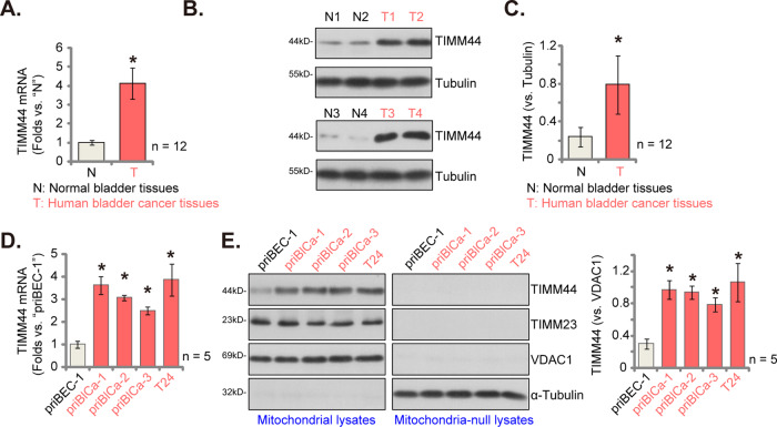
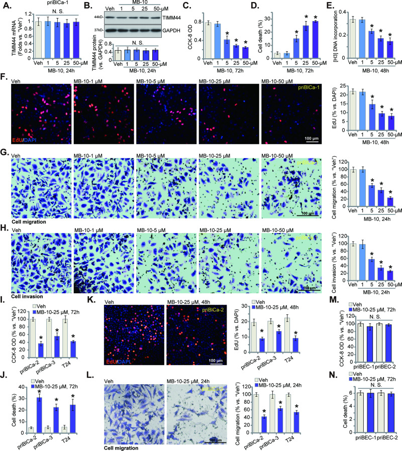
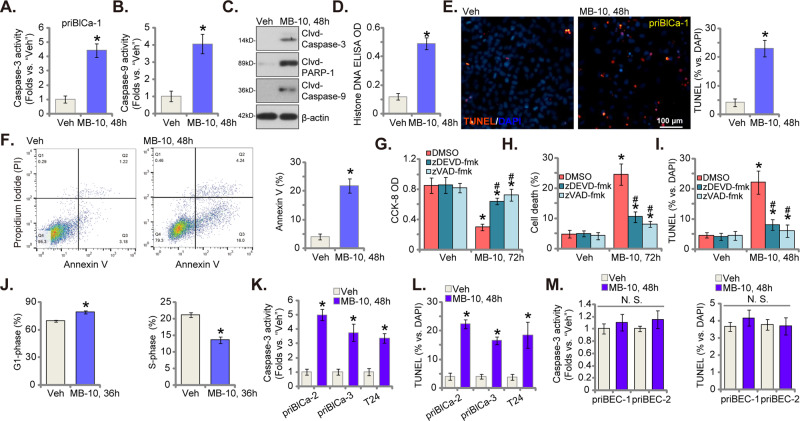
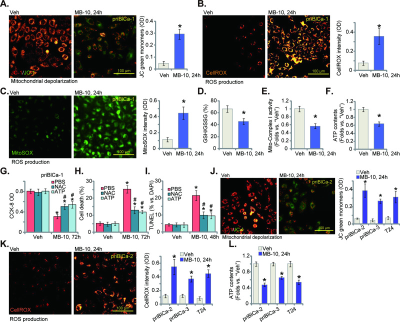
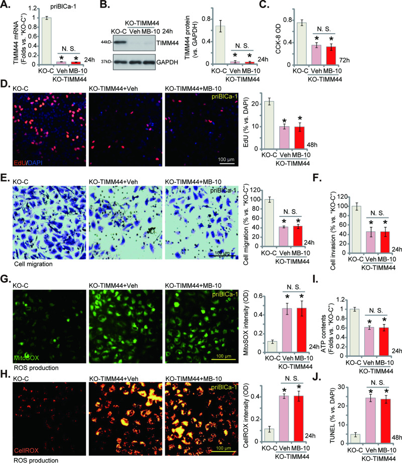
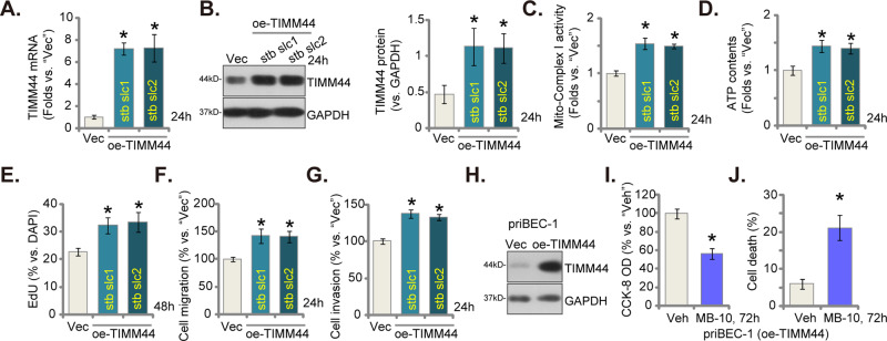
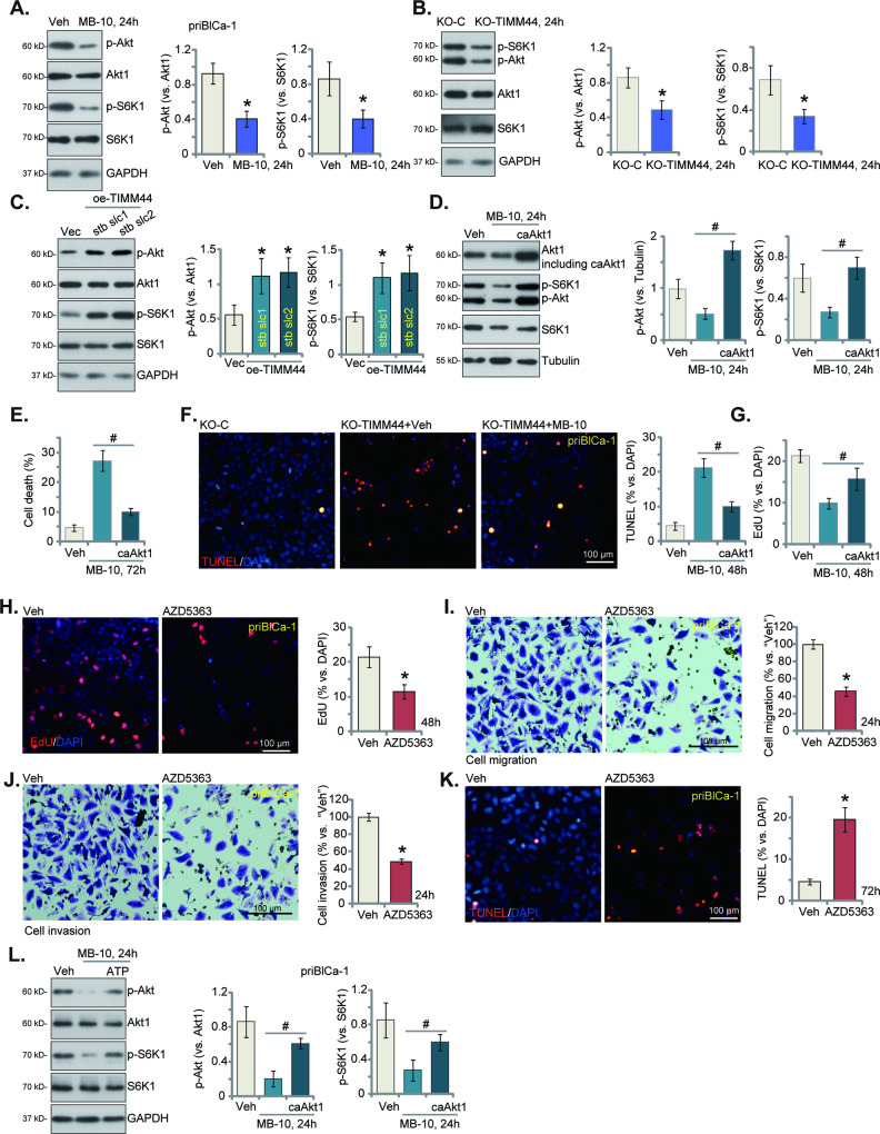
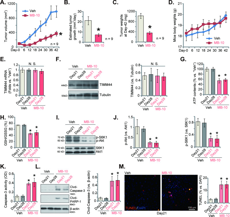
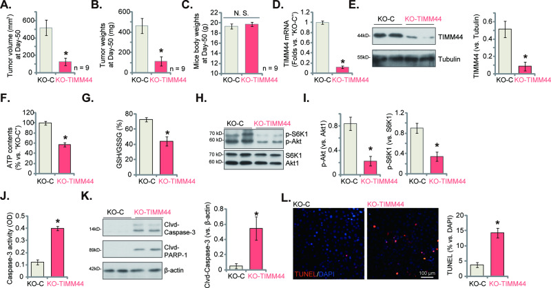
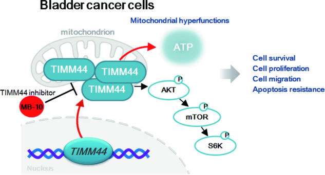
Mitochondria play a multifaceted role in supporting bladder cancer progression. Translocase of inner mitochondrial membrane 44 (TIMM44) is essential for maintaining function and integrity of mitochondria. We here tested the potential effect of MB-10 (MitoBloCK-10), a first-in-class TIMM44 blocker, against bladder cancer cells. TIMM44 mRNA and protein expression is significantly elevated in both human bladder cancer tissues and cells. In both patient-derived primary bladder cancer cells and immortalized (T24) cell line, MB-10 exerted potent anti-cancer activity and inhibited cell viability, proliferation and motility. The TIMM44 blocker induced apoptosis and cell cycle arrest in bladder cancer cells, but failed to provoke cytotoxicity in primary bladder epithelial cells. MB-10 disrupted mitochondrial functions in bladder cancer cells, causing mitochondrial depolarization, oxidative stress and ATP reduction. Whereas exogenously-added ATP and the antioxidant N-Acetyl Cysteine mitigated MB-10-induced cytotoxicity of bladder cancer cells. Genetic depletion of TIMM44 through CRISPR-Cas9 method also induced robust anti-bladder cancer cell activity and MB-10 had no effect in TIMM44-depleted cancer cells. Contrarily, ectopic overexpression of TIMM44 using a lentiviral construct augmented proliferation and motility of primary bladder cancer cells. TIMM44 is important for Akt-mammalian target of rapamycin (mTOR) activation. In primary bladder cancer cells, Akt-S6K1 phosphorylation was decreased by MB-10 treatment or TIMM44 depletion, but enhanced after ectopic TIMM44 overexpression. In vivo, intraperitoneal injection of MB-10 impeded bladder cancer xenograft growth in nude mice. Oxidative stress, ATP reduction, Akt-S6K1 inhibition and apoptosis were detected in MB-10-treated xenograft tissues. Moreover, genetic depletion of TIMM44 also arrested bladder cancer xenograft growth in nude mice, leading to oxidative stress, ATP reduction and Akt-S6K1 inhibition in xenograft tissues. Together, targeting overexpressed TIMM44 by MB-10 significantly inhibits bladder cancer cell growth in vitro and in vivo.


Conflict of interest statement

In this study, all authors affirm that the research was carried out without any commercial or financial associations that might raise concerns about potential conflicts of interest. There are no reported conflicts of interest among the authors.

Figures

Fig. 1
Fig. 1. TIMM44 is overexpression in human bladder cancer tissues and cells.
TIMM44 mRNA (A) and protein (B, C) expression in the described human bladder cancer tissues (“T”) and surrounding normal bladder tissue (“N”) tissues was tested. TIMM44 mRNA expression in both primary (priBlCa-1/priBlCa-2/priBlCa-3) and immortalized (T24) bladder cancer cells as well as in priBEC-1 bladder epithelial cells was shown (D). The mitochondrial fraction lysates and mitochondria-null lysates of the above cells were obtained and expression of listed protein was tested (E). The data were presented as mean ± standard deviation (SD). In (AC), 12 sets of patients tissues were in each group (n = 12). For (D, E) n = 5 stands for five biological repeats. * P < 0.05 vs. “N” tissues/priBEC-1 cells.
Fig. 2
Fig. 2. MB-10 exerts significant anti-bladder cancer cell activity, inhibiting cell viability, proliferation and mobility.
The priBlCa-1 primary bladder cancer cells were maintained under complete medium, treated with MB-10 at designated concentration and cultivated for indicated hours, TIMM44 mRNA and protein expression was tested (A, B). Cell viability and death were tested by CCK-8 (C) and Trypan blue staining (D) assays, respectively. Cell proliferation was measured via [H3] DNA incorporation assay (E) and nuclear EdU staining assay (F), with in vitro cell migration (G) and invasion (H) tested via “Transwell” assays. The primary bladder cancer cells, priBlCa-2 and priBlCa-3 (derived from two other patients) (IL), T24 immortalized cells (IL) or the primary human bladder epithelial cells (priBEC-1 and priBEC-2) (M, N) were maintained under complete medium, treated with MB-10 (25 μM) and cultivated for indicated hours, cell viability (CCK-8 assay, I, M), death (Trypan blue staining assay, J, N), proliferation (nuclear EdU staining assay, K) and migration (“Transwell” assay, L) were tested. “Veh” stands for the vehicle control treatment (0.1% DMSO). The data were presented as mean ± standard deviation (SD). n = 5 stands for five biological repeats. * P < 0.05 vs. “Veh”. “N. S.” stands for non-statistical difference (P > 0.05). The in vitro experiments were repeated five times with similar results obtained. Scale bar = 100 μm.
Fig. 3
Fig. 3. MB-10 induces mitochondrial apoptosis cascade activation in bladder cancer cells.
The priBlCa-1 primary bladder cancer cells were maintained under complete medium, treated with MB-10 (25 μM) and cultivated for indicated hours, Caspase-3 activity (A), Caspase-9 activity (B) and expression of apoptosis-related proteins (C) were tested. Histone-bound DNA contents were measured by ELISA (D); Cell apoptosis was measured via TUNEL-nuclei fluorescence staining (E) and Annexin V-propidium iodide (PI) flow cytometry (F) assays; Cell cycle progression was measured via PI flow cytometry assays (J); The priBlCa-1 primary bladder cancer cells were pretreated for 45 min with the Caspase-3 specific inhibitor zDEVD-fmk (50 μM) or the pan Caspase inhibitor zVAD-fmk (50 μM), followed by MB-10 (25 μM) stimulation, cells were further cultivated for indicated time periods, cell viability, death and apoptosis were tested by CCK-8 (G), Trypan blue staining (H) and TUNEL-nuclei fluorescence staining (I) assays, respectively. The primary bladder cancer cells (priBlCa-2 and priBlCa-3), T24 immortalized cells or the primary human bladder epithelial cells (priBEC-1 and priBEC-2) were maintained under complete medium, treated with MB-10 (25 μM) and cultivated for indicated hours, Caspase-3 activity (K, M) and apoptosis (TUNEL-nuclei fluorescence staining, L, M) were tested. “Veh” stands for the vehicle control treatment (0.1% DMSO). The data were presented as mean ± standard deviation (SD). n = 5 stands for five biological repeats. *P < 0.05 vs. “Veh”. “N. S.” stands for non-statistical difference (P > 0.05). #P < 0.05 vs. “DMSO” pre-treatment (GI). The in vitro experiments were repeated five times with similar results obtained. Scale bar = 100 μm.
Fig. 4
Fig. 4. MB-10 impairs mitochondrial functions in bladder cancer cells.
The priBlCa-1 primary bladder cancer cells were maintained under complete medium, treated with MB-10 (25 μM) and cultivated for indicated hours, mitochondrial depolarization was measured via JC-1 staining assay (A); Mitochondrial ROS production was measured by CellROX fluorescence staining (B) and MitoSOX fluorescence staining (C) assays; GSH/GSSG ratio was also measured (D); The mitochondrial complex I activity (E) and cellular ATP contents (F) were tested as well. The priBlCa-1 cells were pretreated for 45 min with ATP (1 mM) or NAC (400 μM), followed by MB-10 (25 μM) stimulation, cells were further cultivated for indicated time periods, cell viability, death and apoptosis were tested by CCK-8 (G), Trypan blue staining (H) and TUNEL-nuclei fluorescence staining (I) assays, respectively. The primary bladder cancer cells (priBlCa-2 and priBlCa-3) or T24 immortalized cells were maintained under complete medium, treated with MB-10 (25 μM) and cultivated for indicated hours, mitochondrial depolarization was measured via JC-1 staining assay (J); ROS production was measured by CellROX staining assays (K), with cellular ATP contents tested as well (L). “Veh” stands for the vehicle control treatment (0.1% DMSO). The data were presented as mean ± standard deviation (SD). n = 5 stands for five biological repeats. *P < 0.05 vs. “Veh”. #P < 0.05 vs. PBS pretreatment (GI). The in vitro experiments were repeated five times with similar results obtained. Scale bar = 100 μm.
Fig. 5
Fig. 5. TIMM44 knockout exerts significant anti-bladder cancer cell activity.
The priBlCa-1 primary bladder cancer cells with the CRISPR/Cas9-TIMM44-KO construct (“KO-TIMM44”) were treated with or without MB-10 (25 μM) for designated hours; Control cells with the CRISPR/Cas9-KO control construct (“KO-C”) were left untreated. TIMM44 mRNA and protein expression was tested (A, B); Cell viability was tested by CCK-8 assay (C); Cell proliferation was measured via nuclear EdU staining assay (D), and in vitro cell migration (E) and invasion (F) tested via “Transwell” assays; The mitochondrial ROS production was measured by CellROX/MitoSOX fluorescence staining assays (G, H), with ATP contents measured as well (I). Cell apoptosis was tested by TUNEL-nuclei staining assays (J). The data were presented as mean ± standard deviation (SD). n = 5 stands for five biological repeats. *P < 0.05 vs. “KO-C”. “N. S.” stands for non-statistical difference (P > 0.05). The in vitro experiments were repeated five times with similar results obtained. Scale bar = 100 μm.
Fig. 6
Fig. 6. TIMM44 overexpression accelerates proliferation and mobility in bladder cancer cells.
The priBlCa-1 primary bladder cancer cells with the lentiviral TIMM44-overexpressing construct (“oe-TIMM44-stb slc1” and “oe-TIMM44-stb slc2”, representing two stable cell selections) or the empty vector (“Vec”) were cultivated for designated time, TIMM44 mRNA and protein expression was tested (A, B); The mitochondrial complex I activity (C) and cellular ATP contents (D) were measured. Cell proliferation (EdU staining assays, E), in vitro cell migration (F) and invasion (G) were tested as well. TIMM44 protein expression in priBEC-1 bladder epithelial cells with the lentiviral TIMM44-overexpressing construct (“oe-TIMM44”) or the empty vector (“Vec”) was shown (H). The oe-TIMM44 priBEC-1 cells were treated with MB-10 (25 μM) or vehicle control (“Veh”) for indicated hours, cell viability and death were tested by CCK-8 (I) and Trypan blue staining (J) assays, respectively. The data were presented as mean ± standard deviation (SD). n = 5 stands for five biological repeats. *P < 0.05 vs. “Vec”/ “Veh”. The in vitro experiments were repeated five times with similar results obtained.
Fig. 7
Fig. 7. MB-10 inhibits Akt-mTOR activation in bladder cancer cells.
The priBlCa-1 primary bladder cancer cells were maintained under complete medium, treated with MB-10 (25 μM) and cultivated for twenty-four hours, expression of listed proteins was tested (A). The priBlCa-1 primary bladder cancer cells with the CRISPR/Cas9-TIMM44-KO construct (“KO-TIMM44”), the CRISPR/Cas9-KO control construct (“KO-C”), the lentiviral TIMM44-overexpressing construct (“oe-TIMM44-stb slc1” and “oe-TIMM44-stb slc2”, representing two stable cell selections) or the empty vector (“Vec”), were established and maintained under complete medium for twenty-four hours, expression of listed proteins was shown (B, C). The priBlCa-1 primary cells with constitutively-active (S473D) mutant Akt1 (caAkt1) were treated with MB-10 for indicated time periods, expression of listed proteins was shown (D); Cell death, apoptosis and proliferation were measured via Trypan blue staining (E), TUNEL-nuclei staining (F) and EdU-nuclei staining (G) assays, respectively. The priBlCa-1 primary bladder cancer cells were maintained under complete medium, treated with AZD5363 (2.5 μM) and cultivated for indicated hours. Cell proliferation was measured via nuclear EdU staining assay (H), with in vitro cell migration (I) and invasion (J) tested via “Transwell” assays. Cell apoptosis was tested via TUNEL staining assays (K). The priBlCa-1 cells were pretreated for 45 min with ATP (1 mM), followed by MB-10 (25 μM) stimulation, cells were further cultivated for 24 h, and expression of listed proteins was shown (L). “Veh” stands for the vehicle control treatment (0.1% DMSO). The data were presented as mean ± standard deviation (SD). n = 5 stands for five biological repeats. *P < 0.05 vs. “Veh”/“KO-C”/“Vec” cells. #P < 0.05 (DG, L). The in vitro experiments were repeated five times with similar results obtained. Scale bar = 100 μm.
Fig. 8
Fig. 8. Intraperitoneal injection of MB-10 impedes bladder cancer xenograft growth in nude mice.
The priBlCa-1 xenograft-bearing nude mice were intraperitoneally (i.p.) administrated with MB-10 (20 mg/kg body weight, given at every 48 h for a total of three doses) or vehicle control (“Veh”, PBS + Tween 80); The priBlCa-1 xenograft volumes (A) and animal body weights (D) were recorded; The estimated daily tumor growth was calculated (B); At Day-42, all priBlCa-1 xenografts were carefully isolated and weighed (C); Expression of TIMM44 mRNA and listed proteins in the described priBlCa-1 xenograft tissues was tested (E, F, I, J and L). ATP contents (G), GSH/GSSG ratio (H) and Caspase-3 activity (K) in tissue lysates were examined as well. The priBlCa-1 xenograft slides were also subject to fluorescence detection of TUNEL-positive nuclei (M). The data were presented as mean ± standard deviation (SD). In (AD) nine mice were in each group (n = 9). For (EM) five random tissue pieces in each xenograft were tested (n = 5). *P < 0.05 vs. “Veh” group. “N. S.” stands for non-statistical difference (P > 0.05). Scale bar = 100 μm.
Fig. 9
Fig. 9. TIMM44 KO inhibits priBlCa-1 xenograft growth in mice.
The priBlCa-1 primary bladder cancer cells (six million cells per mouse) with the CRISPR/Cas9-TIMM44-KO construct (“KO-TIMM44”) or the CRISPR/Cas9-KO control construct (“KO-C”) were s.c. injected to the nude mice. After 50 days, all priBlCa-1 xenografts of the two groups were isolated and measured (A, B). The animal body weights were recorded (C). Expression of TIMM44 mRNA and listed proteins in priBlCa-1 xenograft tissues was tested (D, E, H, I and K); ATP contents (F), GSH/GSSG ratio (G) and Caspase-3 activity (J) in tissue lysates were examined as well. The priBlCa-1 xenograft slides were also subjected to detection of TUNEL-positive nuclei (L). The data were presented as mean ± standard deviation (SD). In (AC) nine mice were in each group (n = 9). For (DL) five random tissue pieces in each xenograft were tested (n = 5). *P < 0.05 vs. “KO-C” group. Scale bar = 100 μm.
Fig. 10
Fig. 10. The proposed signaling carton of the present study.
TIMM44 overexpression sustains heightened mitochondrial activity and promotes the Akt-mTOR pathway activation, consequently fostering the in vitro and in vivo growth of bladder cancer cells.

Similar articles

Cited by

References

    1. Kamat AM, Hahn NM, Efstathiou JA, Lerner SP, Malmstrom PU, Choi W, et al. Bladder cancer. Lancet. 2016;388:2796–810. doi: 10.1016/S0140-6736(16)30512-8. - DOI - PubMed
    1. Sharma A, Sharma KL, Gupta A, Yadav A, Kumar A. Gallbladder cancer epidemiology, pathogenesis and molecular genetics: recent update. World J Gastroenterol. 2017;23:3978–98. doi: 10.3748/wjg.v23.i22.3978. - DOI - PMC - PubMed
    1. Siegel RL, Wagle NS, Cercek A, Smith RA, Jemal A. Colorectal cancer statistics, 2023. CA: A Cancer J Clin. 2023;73:233–54. - PubMed
    1. Siegel RL, Miller KD, Wagle NS, Jemal A. Cancer statistics, 2023. CA: A Cancer J Clin. 2023;73:17–48. - PubMed
    1. Tran L, Xiao JF, Agarwal N, Duex JE, Theodorescu D. Advances in bladder cancer biology and therapy. Nat Rev Cancer. 2021;21:104–21. doi: 10.1038/s41568-020-00313-1. - DOI - PMC - PubMed

Publication types

Substances