. 2024 Mar 11;10(1):130.
doi: 10.1038/s41420-024-01902-x.

Protein expression of nucleolar protein 12 in the retina and its implication in protection of retina from UV irradiation damage

Affiliations

Protein expression of nucleolar protein 12 in the retina and its implication in protection of retina from UV irradiation damage

Jingtao Liu et al. Cell Death Discov. .

Abstract

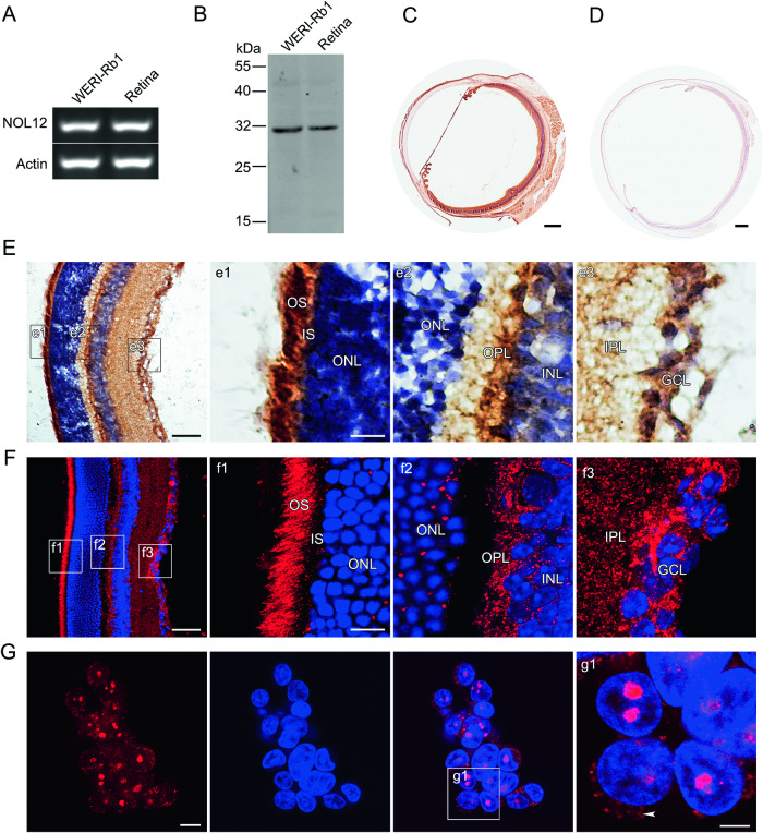
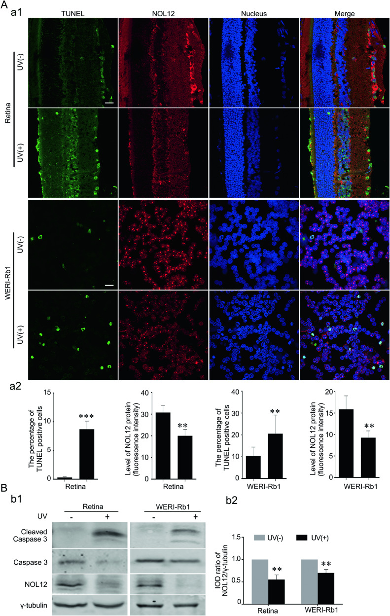
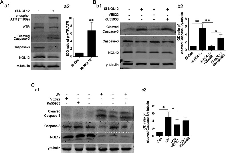
Nucleolar protein 12 (NOL12), one of the nucleolar proteins which are primarily expressed in the nucleolus and play key roles in RNA metabolism, cell proliferation, cell cycle, and cell survival, is widely expressed in various species and multiple organs. Although it has been reported that the mRNA of Drosophila NOL12 homolog viriato is expressed in the eyes of Drosophila, the protein expression of NOL12 in mammalian eyes remains to be elucidated. In this study, we showed through immunohistochemistry that NOL12 was present in the rat retina, with predominant distribution in the cytoplasm of the retinal neuronal cells. In the human retinoblastoma cell line WERI-Rb1, we found that altering NOL12 expression led to a change in WERI-Rb1 cell viability. Knocking down NOL12 expression decreased cell viability. In contrast, overexpressing NOL12 increased cell viability. Furthermore, increasing NOL12 expression inhibited ultraviolet (UV)-induced apoptosis. These findings demonstrated that NOL12 may play an important protective role in retinal cells. In the WERI-Rb1 cells exposed to UV irradiation, we detected that NOL12 was degraded, but this degradation could be attenuated by a pan-Caspase inhibitor. Notably, the inhibitory effect of NOL12 against UV-induced apoptosis could be restrained by increasing the expression of ATR serine/threonine kinase (ATR), a kinase that, when activated by severe DNA damage, can result in apoptosis. We also found that upregulating NOL12 inhibited the activation of ATR caused by UV irradiation. Additionally, inhibiting ATR activity reduced apoptosis resulting from both silencing NOL12 expression and UV exposure. Thus, NOL12 may protect against UV irradiation-induced retinal damage by inhibiting ATR activity.


Conflict of interest statement

The authors declare no competing interests.

Figures

Fig. 1
Fig. 1. NOL12 is mainly expressed in the retinal neuronal cells of the rat eyes.
A, B Expression analyses of NOL12 in adult rat eyes and WERI-Rb1 cells by RT-PCR (A) and Western blot (B). C, D Expression analyses of NOL12 in the adult rat eyes by ABC method of immunohistochemical staining (ABC staining) (C) and the absorption experiment (D). EG Immunohistochemical examination of NOL12 distribution in the retinas of adult rats (E, F) and WERI-Rb1 cells (G) by ABC staining (E) and immunofluorescence staining (F, G); high-power images of the boxed areas in EG are shown in e1-e3, f1-f3, and g1. NOL12 predominantly localizes in the cytoplasm of cells in the retinal nerve layer of adult rats (E, F) but in the nucleus of WERI-Rb1 cells (G); arrowheads indicate the low levels of cytoplasmic localization of NOL12 in WERI-Rb1 cells (g1). A rabbit polyclonal antibody against NOL12 was used in Western blotting (B) and immunohistochemical staining, including ABC staining (CE) and immunofluorescence staining (F, G); NOL12 was labeled by RRX (red), and nuclei were counterstained with Hoechst 33258 (blue). GCL ganglion cell layer, IPL inner plexiform layer, INL inner nuclear layer, OPL outer plexiform layer, ONL outer nuclear layer, IS inner segments, and OS outer segments. Scale bar: 500 μm in (C, D), 50 μm in (E, F), 20 μm in (e1, f1), 10 μm in (G), and 5 μm in (g1).
Fig. 2
Fig. 2. Differential subcellular localization of NOL12 in the rat retinas and WERI-Rb1 cells.
A Double immunofluorescence staining for analyzing subcellular localization of NOL12 in the cells of rat retinal tissue, showing the colocalization of NOL12 with the RGCs marker BRN3B (a1) and the dendrite marker MAP2 (a2), but not with the axonal terminal marker GAP43 (a3). B Double immunofluorescence staining of NOL12 and the nucleolar marker fibrillarin in WERI-Rb1 cells showing that NOL12 is primarily distributed in the nucleolus, with a low level of cytoplasmic distribution (arrowheads). High-power images of the boxed areas in B are shown in b1–b4. A rabbit polyclonal antibody against NOL12 was used in immunofluorescence staining in a1, a3, and B, while a mouse monoclonal antibody against NOL12 was used in immunofluorescence staining in a2. NOL12 was labeled by RRX (red), BRN3B, GAP43, MAP2, and fibrillarin were labeled by FITC (green), and nuclei were counterstained with Hoechst 33258 (blue). RGCs, retinal ganglion cells. Scale bar: 20 μm in A and B, 5 μm in b1.
Fig. 3
Fig. 3. Inhibition of NOL12 expression leads to the apoptosis of retinal cells.
A, B The effect of overexpression of NOL12 (A) and NOL12 silence (B) on the viability of the WERI-Rb1 cell was examined by Alamar blue analysis. C The effect of silencing NOL12 expression with Si-NOL12 on apoptosis in WERI-Rb1 cells was analyzed by TUNEL staining. A rabbit polyclonal antibody against NOL12 was used to stain NOL12 (labeled with RRX [red]), a TUNEL staining kit labeled apoptotic cells (green), and Hoechst 33258 (blue) counterstained the nuclei (c1). The protein level of NOL12 detected by immunofluorescence staining (c2) and the apoptotic rate measured by TUNEL assay (c3) were statistically analyzed. D Flow cytometric analysis of the effect of NOL12 expression inhibition by Si-NOL12 on apoptosis in WERI-Rb1 cells: Annexin V/PI flow cytometry was utilized to determine the apoptotic cells (d1), and the percentage of apoptotic cells was statistically analyzed (d2). E The effect of NOL12 silence on the expression of cleaved Caspase-3 was detected by Western blot analysis using a rabbit polyclonal antibody against NOL12. Representative Western blot of cleaved Caspase-3, Caspase-3, and NOL12 are shown (e1), and the band intensity of cleaved Caspase-3 was analyzed (e2). Data were expressed as mean ± SD. n = 10 for A and B, and n = 3 for CE; *P < 0.05, **P < 0.01. Scale bar, 10 μm.
Fig. 4
Fig. 4. NOL12 protects retinal cells from UV irradiation-induced apoptosis.
A Western blotting was employed to assess the impact of NOL12 overexpression on the expression of cleaved Caspase-3 in WERI-Rb1 cells under UV irradiation. A rabbit polyclonal antibody against NOL12 was used to detect the expression of NOL12 (a1). Representative Western blot of cleaved Caspase-3, Caspase-3, and NOL12 are shown (a1), and the band intensity of cleaved Caspase-3 was analyzed (a2). B Flow cytometry analysis representing the inhibitory effect of NOL12 overexpression on apoptosis in WERI-Rb1 cells under UV irradiation. Apoptotic cells were determined by Annexin V/PI flow cytometry (b1), and the percentages of apoptotic cells was statistically analyzed (b2). Data were expressed as mean ± SD. n = 3. *P < 0.05; **P < 0.01.
Fig. 5
Fig. 5. UV irradiation induces retina injury accompanied by the decrease of NOL12 protein.
A The immunofluorescence and TUNEL double staining revealed apoptosis induced by UV, accompanied by a decrease in NOL12 protein levels in UV-irradiated retinas and WERI-Rb1 cells. A rabbit polyclonal antibody against NOL12 was used to stain NOL12 (labeled with RRX [red]), a TUNEL staining kit labeled apoptotic cells (green), and Hoechst 33258 (blue) counterstained the nuclei (a1); the protein level of NOL12 detected by immunofluorescence staining and the apoptotic rate measured by TUNEL assay were statistically analyzed (a2). B The effect of UV on the expression of cleaved Caspase-3 was detected by Western blot analysis using a rabbit polyclonal antibody against NOL12. Representative Western blots of cleaved Caspase-3, Caspase-3, and NOL12 are shown (b1), and the band intensity of NOL12 was analyzed (b2). Data were expressed as mean ± SD. n = 3. *P < 0.05; **P < 0.01; ***P < 0.001. Scale bar, 20 μm.
Fig. 6
Fig. 6. UV irradiation degrades NOL12 through the Caspase activation-dependent manner.
A Analysis of NOL12 mRNA expression in rat retinas and WERI-Rb1 cells following UV irradiation was conducted using semi-quantitative RT-PCR (a1) and qRT-PCR (a2). B Detection of NOL12 protein expression by Western blotting demonstrates that Z-VAD-FMK inhibits the UV-induced decrease in NOL12 protein level, while Calpeptin does not. Rabbit polyclonal antibody against NOL12 was used in Western blot. A representative Western blot of NOL12 is shown (b1), and the band intensity was analyzed (b2). Z-VAD-FMK, the inhibitor of pan-Caspase kinases. Calpeptin, the inhibitor of pan-calpain kinases. Data were expressed as mean ± SD. n = 3. *P < 0.05; **P < 0.01.
Fig. 7
Fig. 7. NOL12 interacts with ATR in the rat retina and WERI-Rb1 cells.
A Double immunofluorescence staining of NOL12 and ATR in WERI-Rb1 cells showing that NOL12 is well colocalized with ATR in WERI-Rb1 cells, predominantly in the nucleoli (arrowheads) and to a lesser extent in the cytoplasm (arrows). High-power images of the boxed areas in (A) are shown in a1–a8. B The interactions between NOL12, ATR, and its substrate RPA in WERI-Rb1 cells were detected by co-immunoprecipitation assays. C Double immunofluorescence staining of NOL12 and ATR in adult rat retinas showing that NOL12 is well colocalized with ATR in the cytoplasm of RGCs. High-power images of the boxed areas in (C) are shown in c1–c4. D The interactions between NOL12, ATR, and its substrate RPA in adult rat retinas were detected by co-immunoprecipitation assays. Rabbit polyclonal antibody against NOL12 and mouse monoclonal antibody against ATR was utilized in immunofluorescence staining (A, C). Rabbit polyclonal antibodies against NOL12 and ATR were utilized in Western blot (B, D). NOL12 is labeled by RRX (red), ATR by FITC (green), and nuclei are counterstained with Hoechst 33258 (blue) (A, C). Scale bars: 5 μm in A and 10 μm in C.
Fig. 8
Fig. 8. NOL12 inhibits the activation of ATR to protect against UV irradiation-induced apoptosis.
A The expression of cleaved Caspase-3 and the activation of ATR were detected by Western blotting, showing that NOL12 overexpression inhibits the ATR activation and apoptosis induced by UV irradiation, while, ATR overexpression reverses the inhibitory role of NOL12 on the apoptosis. Rabbit polyclonal antibodies against NOL12 and ATR were utilized in Western blot. Representative Western blot bands are shown (a1), and the band intensities of p-ATR (a2) and cleaved Caspase-3 (a3) were analyzed. B Flow cytometric analysis was conducted to assess the impact of ATR overexpression on the inhibitory role of NOL12 in UV irradiation-induced apoptosis in WERI-Rb1 cells: Annexin V/PI flow cytometry was utilized to determine the apoptotic cells (b1), and the percentages of apoptotic cells underwent statistical analysis (b2). Data were expressed as mean ± SD. n = 3. *P < 0.05; **P < 0.01.
Fig. 9
Fig. 9. Inhibitor of ATR blocks the apoptosis induced by UV irradiation and NOL12 depletion.
A The effect of silencing NOL12 expression with Si-NOL12 on the activation of ATR in WERI-Rb1 cells was analyzed by Western Blotting. B, C Detection of expression levels of cleaved Caspase-3 by Western blotting demonstrating that VE822 but not KU55933 inhibited apoptosis induced by NOL12 silencing (B) and UV irradiation (C). Rabbit polyclonal antibody against NOL12 and rabbit polyclonal antibody against ATR were utilized in Western blot. Representative Western blot bands were shown (a1, b1, and c1), and the band intensities were analyzed (a2, b2, and c2). Data were expressed as mean ± SD. n = 3. *P < 0.05, **P < 0.01.

Similar articles

Cited by

References

    1. Andersen JS, Lam YW, Leung AK, Ong SE, Lyon CE, Lamond AI, et al. Nucleolar proteome dynamics. Nature. 2005;433:77–83. doi: 10.1038/nature03207. - DOI - PubMed
    1. Ogawa LM, Baserga SJ. Crosstalk between the nucleolus and the DNA damage response. Mol Biosyst. 2017;13:443–55. doi: 10.1039/C6MB00740F. - DOI - PMC - PubMed
    1. Spriggs KA, Bushell M, Willis AE. Translational regulation of gene expression during conditions of cell stress. Mol cell. 2010;40:228–37. doi: 10.1016/j.molcel.2010.09.028. - DOI - PubMed
    1. van Sluis M, McStay B. Nucleolar reorganization in response to rDNA damage. Curr Opin Cell Biol. 2017;46:81–6. doi: 10.1016/j.ceb.2017.03.004. - DOI - PubMed
    1. Suzuki S, Kanno M, Fujiwara T, Sugiyama H, Yokoyama A, Takahashi H, et al. Molecular cloning and characterization of Nop25, a novel nucleolar RNA binding protein, highly conserved in vertebrate species. Exp Cell Res. 2006;312:1031–41. doi: 10.1016/j.yexcr.2005.12.017. - DOI - PubMed