Review
. 2024 Mar 11;15(3):202.
doi: 10.1038/s41419-024-06559-z.

Cellular heterogeneity in TNF/TNFR1 signalling: live cell imaging of cell fate decisions in single cells

Affiliations
Review

Cellular heterogeneity in TNF/TNFR1 signalling: live cell imaging of cell fate decisions in single cells

Marcus K Preedy et al. Cell Death Dis. .

Abstract

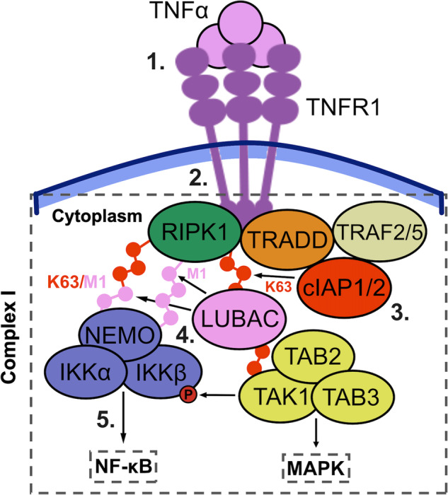
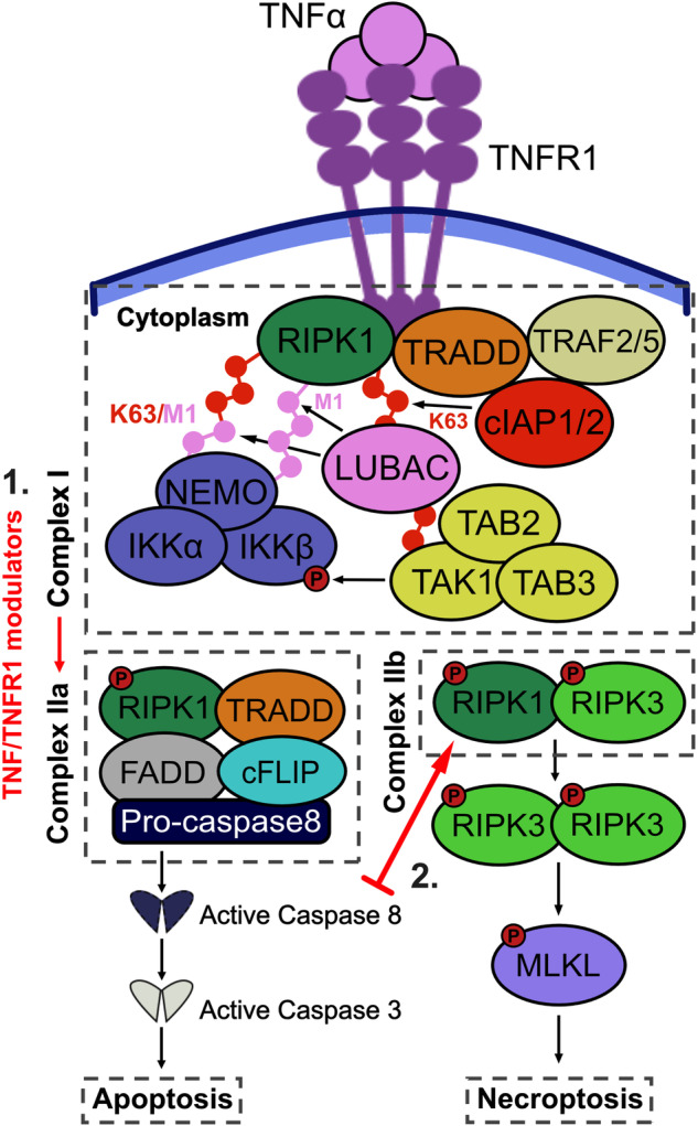
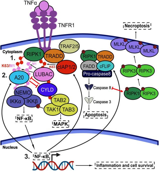
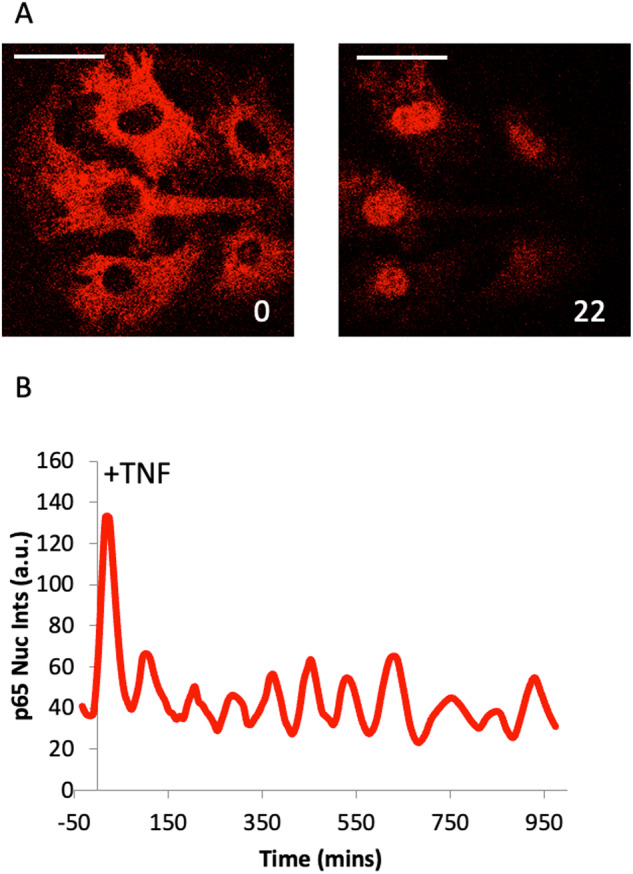
Cellular responses to TNF are inherently heterogeneous within an isogenic cell population and across different cell types. TNF promotes cell survival by activating pro-inflammatory NF-κB and MAPK signalling pathways but may also trigger apoptosis and necroptosis. Following TNF stimulation, the fate of individual cells is governed by the balance of pro-survival and pro-apoptotic signalling pathways. To elucidate the molecular mechanisms driving heterogenous responses to TNF, quantifying TNF/TNFR1 signalling at the single-cell level is crucial. Fluorescence live-cell imaging techniques offer real-time, dynamic insights into molecular processes in single cells, allowing for detection of rapid and transient changes, as well as identification of subpopulations, that are likely to be missed with traditional endpoint assays. Whilst fluorescence live-cell imaging has been employed extensively to investigate TNF-induced inflammation and TNF-induced cell death, it has been underutilised in studying the role of TNF/TNFR1 signalling pathway crosstalk in guiding cell-fate decisions in single cells. Here, we outline the various opportunities for pathway crosstalk during TNF/TNFR1 signalling and how these interactions may govern heterogenous responses to TNF. We also advocate for the use of live-cell imaging techniques to elucidate the molecular processes driving cell-to-cell variability in single cells. Understanding and overcoming cellular heterogeneity in response to TNF and modulators of the TNF/TNFR1 signalling pathway could lead to the development of targeted therapies for various diseases associated with aberrant TNF/TNFR1 signalling, such as rheumatoid arthritis, metabolic syndrome, and cancer.


Conflict of interest statement

The authors declare no competing interests.

Figures

Fig. 1
Fig. 1. TNF/TNFR1 signalling through complex I.
1 Trimeric TNF binds to TNFR1 on the cell-surface membrane of target cells and induces oligomerization of the receptor. 2 TRADD and RIPK1 are recruited to the intracellular domains of TNF/TNFR1 through their ‘death domain’. These proteins then recruit TRAF2/5 and cIAP1/2 to form TNF/TNFR1 complex I. 3 cIAP1/2 adds K63-linked ubiquitin chains to RIPK1, allowing for the recruitment of LUBAC and TAB2/3. TAB2/3 recruits TAB1 and TAK1. TAK1 then activates the MAPK signalling pathway. 4 LUBAC adds M1-linked ubiquitin chains to RIPK1 and potentially generates K63/M1-linked hybrids. M1- and K63/M1- linked ubiquitin chains on RIPK1 allow for the recruitment of the IKK complex through NEMO. Recruitment of the IKK complex to TNFR1 brings it in proximity to TAK1, which phosphorylates and activates IKK2. 5 IKK2 phosphorylates IκBα, leading to its ubiquitin-mediated proteasomal degradation and liberation of NF-κB, thus activating the NF-κB signalling pathway.
Fig. 2
Fig. 2. TNF/TNFR1 signalling through complex II.
TNF stimulation predominantly induces the formation of TNF/TNFR1 complex I, leading to activation of pro-inflammatory NF-κB and MAPK signalling pathways. However, TNF/TNFR1 signalling may also trigger apoptosis and necroptosis via complex IIa and IIb, respectively. 1 TNF/TNFR1-disrupting agents, such as SMAC mimetics and TAK1 inhibitors, can promote the dissociation of complex I and formation of complex II. In complex IIa, RIPK1 associates with TRADD, FADD, cFLIP, and pro-caspase 8. The high local concentration of pro-caspase 8 induces caspase 8 activation through autocleavage and trans-cleavage from other active caspases. Caspase 8 then cleaves and activates caspase 3, which in turn cleaves downstream components to induce apoptosis. 2 If caspase activation is inhibited, complex IIb can prevail as the dominant signalling pathway. In this pathway, RIPK1 is phosphorylated by RIPK3, causing it to dissociate from complex IIa and instead form a RIPK1-RIPK3 pro-necrotic complex. RIPK3 then directly phosphorylates MLKL, causing it to oligomerise and translocate to the plasma membrane. MLKL binds to phosphatidylinositol phosphates on the membrane’s inner leaflet and disrupts the integrity of the cell membrane, resulting in the release of intracellular contents. This process leads to cell swelling, rupture, and ultimately culminates in cell death by necroptosis.
Fig. 3
Fig. 3. Interplay between TNF/TNFR1 complex I and complex II signalling pathways.
TNF stimulation activates a series of complex signalling cascades that drive cell-fate decisions. The intricate interplay between these signalling pathways plays a pivotal role in determining a cell’s response to TNF. There are several checkpoints throughout TNF/TNFR1 signalling where crosstalk can occur. 1 One of the primary components believed to govern the interplay between TNF/TNFR1 complex I- and complex II-mediated signalling occurs during the early stages of complex formation. The post-translational modification (PTM) profile of RIPK1 determines whether signalling will predominantly propagate from complex I or complex II [97]. Under specific conditions, RIPK1 can experience significant PTM alterations, causing it to dissociate from TNF/TNFR1 complex I and instead form complex IIa. 2 The PTM profile of RIPK1 is thought to be regulated by the antagonistic interactions of A20 and CYLD. A20 binds to M1-linked chains on RIPK1 and protects them from degradation by CYLD, thus stabilising complex I. If CYLD successfully removes M1-linked ubiquitin, RIPK1 is more likely to dissociate from complex I and form complex IIa. 3 TNF/TNFR1 complex I-driven activation of NF-κB leads to increased expression of pro-survival genes, such as cIAP1/2 and c-FLIP. c-FLIP directly inhibits activation of pro-caspase 8, thus reducing signalling downstream of complex IIa. A20 is also under NF-κB transcriptional control, adding another layer of complexity to TNF/TNFR1 signalling crosstalk.
Fig. 4
Fig. 4. NF-κB oscillations in single cells.
Mouse ear fibroblasts from an eGFP-RelA Bacterial Artificial Chromosome transgenic line were imaged on a Zeiss LSM780 confocal microscope every 2 min for 16 h following 10 ng/ml TNF stimulation. A Images at 0 min and 22 min after TNF stimulation (scale bar 10 microns) (B) Analysis of nuclear fluorescence in an example single-cell over time.

Similar articles

Cited by

References

    1. Tartaglia LA, Weber RF, Figari IS, Reynolds C, Palladino MA, Jr, Goeddel DV. The two different receptors for tumor necrosis factor mediate distinct cellular responses. Proc Natl Acad Sci USA. 1991;88:9292–6. doi: 10.1073/pnas.88.20.9292. - DOI - PMC - PubMed
    1. Sedger LM, McDermott MF. TNF and TNF-receptors: from mediators of cell death and inflammation to therapeutic giants - past, present and future. Cytokine Growth Factor Rev. 2014;25:453–72. doi: 10.1016/j.cytogfr.2014.07.016. - DOI - PubMed
    1. Probert L. TNF and its receptors in the CNS: the essential, the desirable and the deleterious effects. Neuroscience. 2015;302:2–22. doi: 10.1016/j.neuroscience.2015.06.038. - DOI - PubMed
    1. Bradley JR. TNF-mediated inflammatory disease. J Pathol. 2008;214:149–60. doi: 10.1002/path.2287. - DOI - PubMed
    1. Beldi G, Bahiraii S, Lezin C, Nouri Barkestani M, Abdelgawad ME, Uzan G, et al. TNFR2 is a crucial hub controlling mesenchymal stem cell biological and functional properties. Front Cell Dev Biol. 2020;8:596831. doi: 10.3389/fcell.2020.596831. - DOI - PMC - PubMed

Publication types

MeSH terms

Substances