

Bone scan findings of Paget's disease of bone in patients with VCP Multisystem Proteinopathy 1
- PMID: 38467645
- PMCID: PMC10928154
- DOI: 10.1038/s41598-024-54526-7
Bone scan findings of Paget's disease of bone in patients with VCP Multisystem Proteinopathy 1
Abstract
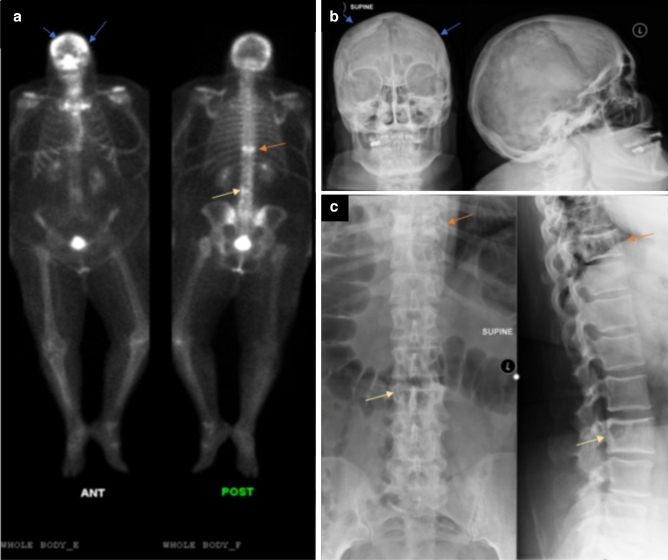
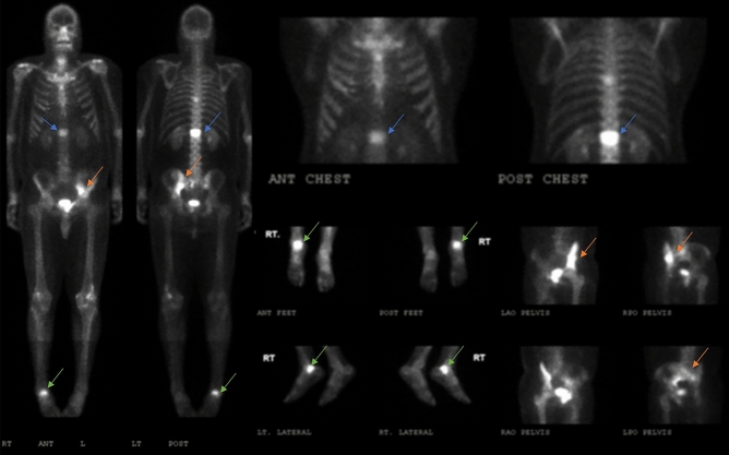
Multisystem Proteinopathy 1 (MSP1) disease is a rare genetic disorder caused by mutations in the Valosin-Containing Protein (VCP) gene with clinical features of inclusion body myopathy (IBM), frontotemporal dementia (FTD), and Paget's disease of bone (PDB). We performed bone scan imaging in twelve patients (6 females, 6 males) with confirmed VCP gene mutation six (50%) of which has myopathy alone, four (33%) with both PDB and myopathy, and two (15%) were presymptomatic carriers. We aim to characterize the PDB in diagnosed individuals, and potentially identify PDB in the myopathy and presymptomatic groups. Interestingly, two patients with previously undiagnosed PDB had positive diagnostic findings on the bone scan and subsequent radiograph imaging. Among the individuals with PDB, increased radiotracer uptake of the affected bones were of typical distribution as seen in conventional PDB and those reported in other MSP1 cohorts which are the thoracic spine and ribs (75%), pelvis (75%), shoulder (75%) and calvarium (15%). Overall, we show that technetium-99m bone scans done at regular intervals are a sensitive screening tool in patients with MSP1 associated VCP variants at risk for PDB. However, diagnostic confirmation should be coupled with clinical history, biochemical analysis, and skeletal radiographs to facilitate early treatment and prevention complications, acknowledging its limited specificity.
© 2024. The Author(s).
Conflict of interest statement
The authors declare no competing interests.
Figures


Similar articles
-
Cross-sectional study of patients with VCP multisystem proteinopathy 1 using dual-energy x-ray absorptiometry.Muscle Nerve. 2024 Jun;69(6):699-707. doi: 10.1002/mus.28095. Epub 2024 Mar 29. Muscle Nerve. 2024. PMID: 38551101 Free PMC article.
-
Characteristics of VCP mutation-associated cardiomyopathy.Neuromuscul Disord. 2021 Aug;31(8):701-705. doi: 10.1016/j.nmd.2021.06.005. Epub 2021 Jun 12. Neuromuscul Disord. 2021. PMID: 34244020
-
Novel Variants in the VCP Gene Causing Multisystem Proteinopathy 1.Genes (Basel). 2023 Mar 8;14(3):676. doi: 10.3390/genes14030676. Genes (Basel). 2023. PMID: 36980948 Free PMC article.
-
The Cure VCP Scientific Conference 2021: Molecular and clinical insights into neurodegeneration and myopathy linked to multisystem proteinopathy-1 (MSP-1).Neurobiol Dis. 2022 Jul;169:105722. doi: 10.1016/j.nbd.2022.105722. Epub 2022 Apr 8. Neurobiol Dis. 2022. PMID: 35405261 Free PMC article. Review.
-
Sex influences clinical phenotype in valosin-containing protein mutations: A case family report and systematic literature review.Clin Neurol Neurosurg. 2023 Sep;232:107875. doi: 10.1016/j.clineuro.2023.107875. Epub 2023 Jul 5. Clin Neurol Neurosurg. 2023. PMID: 37441929
Cited by
-
2024 VCP International Conference: Exploring multi-disciplinary approaches from basic science of valosin containing protein, an AAA+ ATPase protein, to the therapeutic advancement for VCP-associated multisystem proteinopathy.Neurobiol Dis. 2025 Apr;207:106861. doi: 10.1016/j.nbd.2025.106861. Epub 2025 Mar 2. Neurobiol Dis. 2025. PMID: 40037468 Review.
References
-
- Kimonis, V. Inclusion body myopathy with paget disease of bone and/or frontotemporal dementia. In: GeneReviews® (eds Adam, M. P., Ardinger, H. H., Pagon, R. A. et al.) (University of Washington, Seattle, 1993). Accessed 19 Jul 2021. http://www.ncbi.nlm.nih.gov/books/NBK1476/
MeSH terms
Substances
Supplementary concepts
Grants and funding
LinkOut - more resources
Full Text Sources
Medical
Miscellaneous
