

Point-of-care lung ultrasound predicts hyperferritinemia and hospitalization, but not elevated troponin in SARS-CoV-2 viral pneumonitis in children
- PMID: 38467670
- PMCID: PMC10928070
- DOI: 10.1038/s41598-024-55590-9
Point-of-care lung ultrasound predicts hyperferritinemia and hospitalization, but not elevated troponin in SARS-CoV-2 viral pneumonitis in children
Abstract
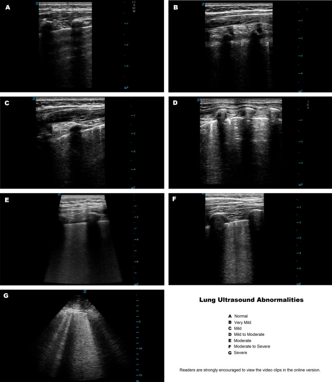
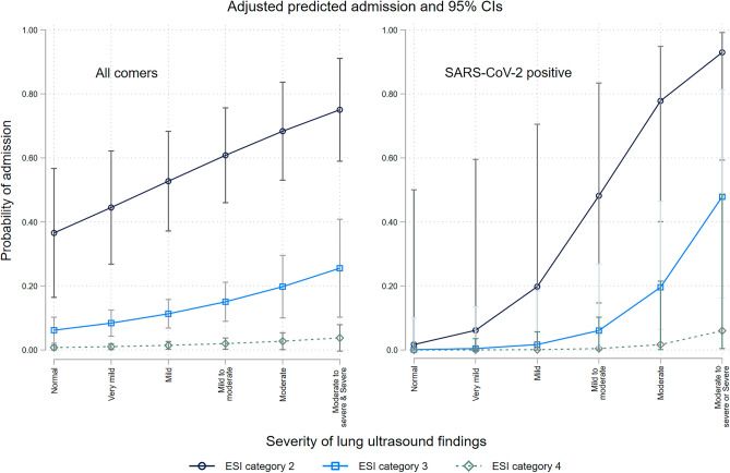
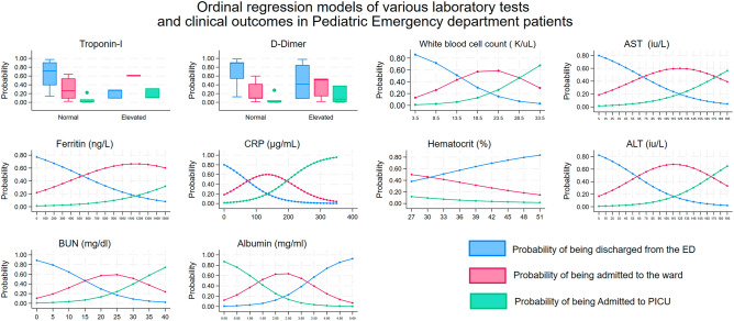
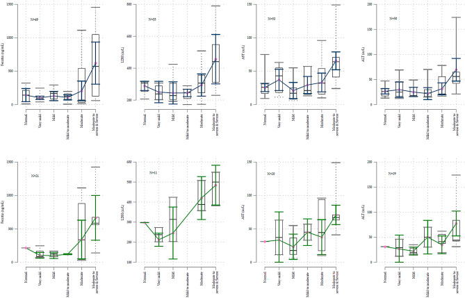
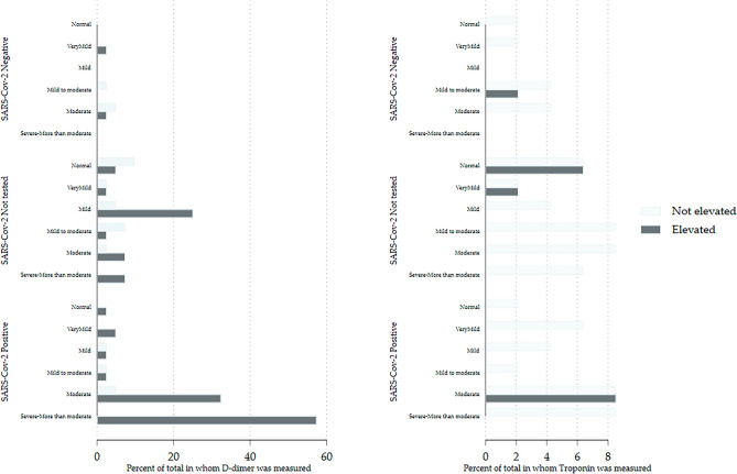
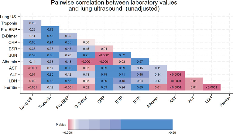
SARS-CoV-2 often causes viral pneumonitis, hyperferritinemia, elevations in D-dimer, lactate dehydrogenase (LDH), transaminases, troponin, CRP, and other inflammatory markers. Lung ultrasound is increasingly used to diagnose and stratify viral pneumonitis severity. We retrospectively reviewed 427 visits in patients aged 14 days to 21 years who had had a point-of-care lung ultrasound in our pediatric emergency department from 30/November/2019 to 14/August/2021. Lung ultrasounds were categorized using a 6-point ordinal scale. Lung ultrasound abnormalities predicted increased hospitalization with a threshold effect. Increasingly abnormal laboratory values were associated with decreased discharge from the ED and increased admission to the ward and ICU. Among patients SARS-CoV-2 positive patients ferritin, LDH, and transaminases, but not CRP or troponin were significantly associated with abnormalities on lung ultrasound and also with threshold effects. This effect was not demonstrated in SARS-CoV-2 negative patients. D-Dimer, CRP, and troponin were sometimes elevated even when the lung ultrasound was normal.
© 2024. The Author(s).
Conflict of interest statement
The authors declare no competing interests.
Figures
Similar articles
-
Diagnostic yield of point-of-care ultrasound imaging of the lung in patients with COVID-19.Emergencias. 2020 Sep;32(5):340-344. Emergencias. 2020. PMID: 33006834 English, Spanish.
-
The role of lung ultrasound as a frontline diagnostic tool in the era of COVID-19 outbreak.Intern Emerg Med. 2021 Apr;16(3):749-756. doi: 10.1007/s11739-020-02524-8. Epub 2020 Oct 22. Intern Emerg Med. 2021. PMID: 33090353 Free PMC article.
-
Point-of-Care Lung Ultrasound for COVID-19: Findings and Prognostic Implications From 105 Consecutive Patients.J Intensive Care Med. 2021 Mar;36(3):334-342. doi: 10.1177/0885066620988831. J Intensive Care Med. 2021. PMID: 33535883 Free PMC article.
-
Thoracic imaging tests for the diagnosis of COVID-19.Cochrane Database Syst Rev. 2020 Sep 30;9:CD013639. doi: 10.1002/14651858.CD013639.pub2. Cochrane Database Syst Rev. 2020. Update in: Cochrane Database Syst Rev. 2020 Nov 26;11:CD013639. doi: 10.1002/14651858.CD013639.pub3. PMID: 32997361 Updated.
-
Point-of-care lung ultrasound in patients with COVID-19 - a narrative review.Anaesthesia. 2020 Aug;75(8):1096-1104. doi: 10.1111/anae.15082. Epub 2020 Apr 28. Anaesthesia. 2020. PMID: 32275766 Free PMC article. Review.
References
MeSH terms
Substances
Grants and funding
LinkOut - more resources
Full Text Sources
Medical
Research Materials
Miscellaneous
