. 2024 Mar 11;10(1):54.
doi: 10.1038/s41531-024-00670-w.

Disrupted sleep-wake regulation in the MCI-Park mouse model of Parkinson's disease

Affiliations

Disrupted sleep-wake regulation in the MCI-Park mouse model of Parkinson's disease

K C Summa et al. NPJ Parkinsons Dis. .

Erratum in

Abstract

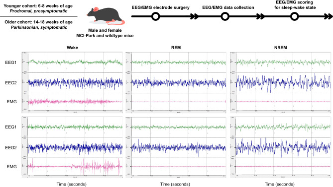
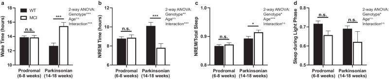
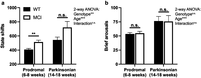
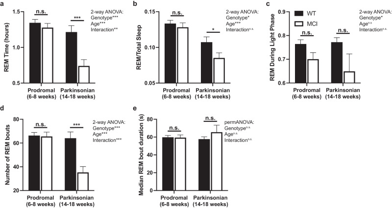
Disrupted sleep has a profound adverse impact on lives of Parkinson's disease (PD) patients and their caregivers. Sleep disturbances are exceedingly common in PD, with substantial heterogeneity in type, timing, and severity. Among the most common sleep-related symptoms reported by PD patients are insomnia, excessive daytime sleepiness, and sleep fragmentation, characterized by interruptions and decreased continuity of sleep. Alterations in brain wave activity, as measured on the electroencephalogram (EEG), also occur in PD, with changes in the pattern and relative contributions of different frequency bands of the EEG spectrum to overall EEG activity in different vigilance states consistently observed. The mechanisms underlying these PD-associated sleep-wake abnormalities are poorly understood, and they are ineffectively treated by conventional PD therapies. To help fill this gap in knowledge, a new progressive model of PD - the MCI-Park mouse - was studied. Near the transition to the parkinsonian state, these mice exhibited significantly altered sleep-wake regulation, including increased wakefulness, decreased non-rapid eye movement (NREM) sleep, increased sleep fragmentation, reduced rapid eye movement (REM) sleep, and altered EEG activity patterns. These sleep-wake abnormalities resemble those identified in PD patients. Thus, this model may help elucidate the circuit mechanisms underlying sleep disruption in PD and identify targets for novel therapeutic approaches.


Conflict of interest statement

The authors declare no competing interests.

Figures

Fig. 1
Fig. 1. Experimental protocol.
Male and female MCI-Park mice and wildtype littermates were obtained from breeding colonies maintained at Northwestern University and the University of California, Berkeley. Mice ranging from approximately 37–57 days of age (younger cohort; presymptomatic or prodromal MCI-Park mice and age-matched wildtype littermates) and from approximately 88–121 days of age (older cohort; symptomatic parkinsonian MCI-Park mice and age-matched wildtype littermates) underwent electroencephalography (EEG) and electromyography (EMG) recording electrode implantation surgery and a minimum of 7 days of undisturbed recovery. EEG/EMG data were collected continuously and scored as wake, rapid eye movement (REM) sleep, or non-REM (NREM) sleep. Representative 10-s epochs of each vigilance state are depicted to demonstrate characteristic EEG/EMG profiles. EEG1, EEG2, and EMG are all used to classify the vigilance state of each epoch, with EEG2 as the primary electrode used for sleep state classification and power spectral analysis. Epochs in which a state could not be assigned were scored as artifact and excluded from sleep-wake trait analysis. Animals in which the EEG2 signal was poor, excessively noisy, and/or ambiguous as to the vigilance state were excluded from power spectral analysis. Wake exhibits low amplitude high-frequency EEG waves with variable EMG activity. REM sleep exhibits low amplitude high-frequency EEG waves with absent EMG activity. NREM sleep exhibits high amplitude low-frequency EEG waves with absent EMG activity. Descriptions and definitions of each sleep-wake trait examined are provided in the Supplemental Material.
Fig. 2
Fig. 2. Wake and NREM sleep amounts and proportion of total sleep during the light phase.
a Total time spent awake over 24 h in wildtype (filled bars) and MCI-Park mice (open bars) at 6–8 weeks of age and 14–18 weeks of age. b Total time spent in NREM sleep over 24 h in wildtype and MCI-Park mice at 6–8 weeks of age (filled bars) and 14–18 weeks of age (open bars). c Proportion of total sleep (NREM plus REM sleep) that is NREM sleep (NREM sleep amount/total sleep amount) in wildtype (filled bars) and MCI-Park (open bars) mice at 6–8 weeks of age (solid bars) and 14–18 weeks of age (open bars). d Proportion of total sleep during light phase of the light-dark cycle. *p < 0.05; **p < 0.01; ***p < 0.001. Error bars depict standard error of the mean (s.e.m.). N = 10–24 mice per genotype per age. All data presented in this figure were collected at Northwestern University.
Fig. 3
Fig. 3. Wake and NREM bout number and median bout duration.
Number of wake (a) and NREM (c) bouts over 24 h in wildtype (filled bars) and MCI-Park mice (open bars) at 6–8 weeks of age and 14–18 weeks of age. Median wake (b) and NREM (d) bout duration in wildtype (filled bars) and MCI-Park (open bars) mice at 6–8 weeks of age and 14–18 weeks of age. *p < 0.05; **p < 0.01; ***p < 0.001. Error bars depict standard error of the mean (s.e.m.). N = 10–24 mice per genotype per age. All data presented in this figure were collected at Northwestern University.
Fig. 4
Fig. 4. State shifts and brief arousals.
a Number of state shifts over 24 h in wildtype (filled bars) and MCI-Park (open bars) mice at 6–8 weeks of age and 14–18 weeks of age. b Number of brief arousals over 24 h in wildtype (filled bars) and MCI-Park (open bars) mice at 6–8 weeks of age and 14–18 weeks of age. *p < 0.05; **p < 0.01; ***p < 0.001. Error bars depict standard error of the mean (s.e.m.). N = 10–24 mice per genotype per age. All data presented in this figure were collected at Northwestern University.
Fig. 5
Fig. 5. REM sleep amount, distribution, bout number, and median bout duration.
a Total amount of REM sleep over 24 h in wildtype (filled bars) and MCI-Park (open bars) mice at 6–8 weeks of age and 14–18 weeks of age. b Proportion of total sleep that is REM (REM sleep amount/total sleep amount) in wildtype (filled bars) and MCI-Park (open bars) mice at 6–8 weeks of age (solid bars) and 14–18 weeks of age (open bars). c Proportion of REM sleep during the light phase of the light-dark cycle in wildtype (filled bars) and MCI-Park (open bars) mice at 6–8 weeks of age (solid bars) and 14–18 weeks of age (open bars). d Number of REM bouts over 24 h in wildtype (filled bars) and MCI-Park (open bars) mice at 6–8 weeks of age and 14–18 weeks of age. e Median duration of REM bouts in wildtype (filled bars) and MCI-Park (open bars) mice at 6–8 weeks of age and 14–18 weeks of age. *p < 0.05; **p < 0.01; ***p < 0.001. Error bars depict standard error of the mean (s.e.m.). N = 10–24 mice per genotype per age. All data presented in this figure were collected at Northwestern University.

Similar articles

Cited by

References

    1. Poewe W, et al. Parkinson disease. Nat. Rev. Dis. Primers. 2017;3:17013. doi: 10.1038/nrdp.2017.13. - DOI - PubMed
    1. Stefani A, Hogl B. Sleep in Parkinson’s disease. Neuropsychopharmacology. 2020;45:121–128. doi: 10.1038/s41386-019-0448-y. - DOI - PMC - PubMed
    1. Chahine LM, Amara AW, Videnovic A. A systematic review of the literature on disorders of sleep and wakefulness in Parkinson’s disease from 2005 to 2015. Sleep. Med. Rev. 2017;35:33–50. doi: 10.1016/j.smrv.2016.08.001. - DOI - PMC - PubMed
    1. French IT, Muthusamy KA. A Review of Sleep and Its Disorders in Patients with Parkinson’s Disease in Relation to Various Brain Structures. Front. Aging Neurosci. 2016;8:114. doi: 10.3389/fnagi.2016.00114. - DOI - PMC - PubMed
    1. Gros P, Videnovic A. Sleep and Circadian Rhythm Disorders in Parkinson’s Disease. Curr. Sleep Med. Rep. 2017;3:222–234. doi: 10.1007/s40675-017-0079-y. - DOI - PMC - PubMed