. 2024 Mar 11;14(1):5941.
doi: 10.1038/s41598-024-56680-4.

Ascorbic acid mitigates the impact of oxidative stress in a human model of febrile seizure and mesial temporal lobe epilepsy

Affiliations

Ascorbic acid mitigates the impact of oxidative stress in a human model of febrile seizure and mesial temporal lobe epilepsy

Stefania Scalise et al. Sci Rep. .

Abstract

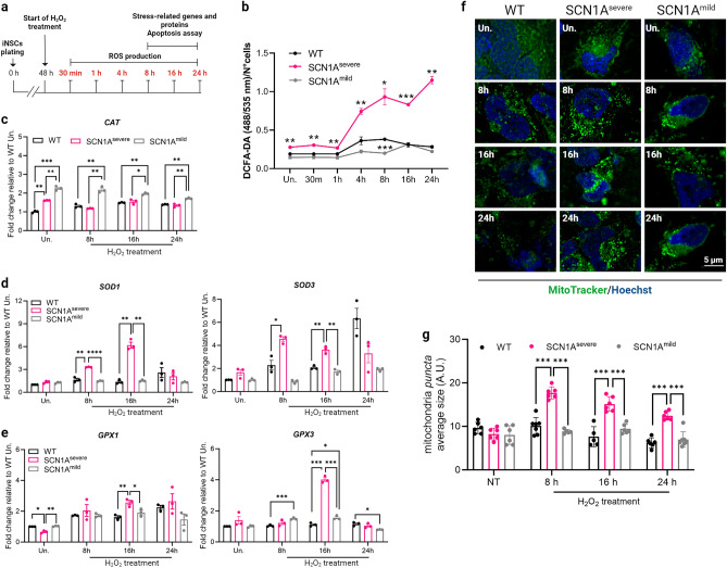
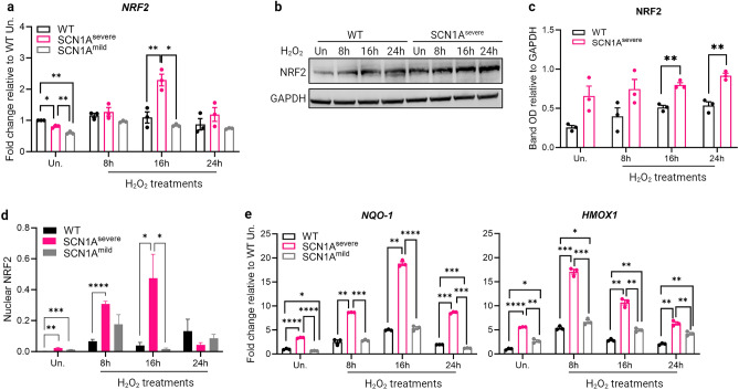
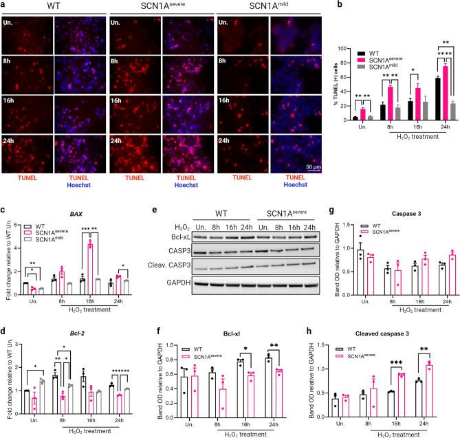
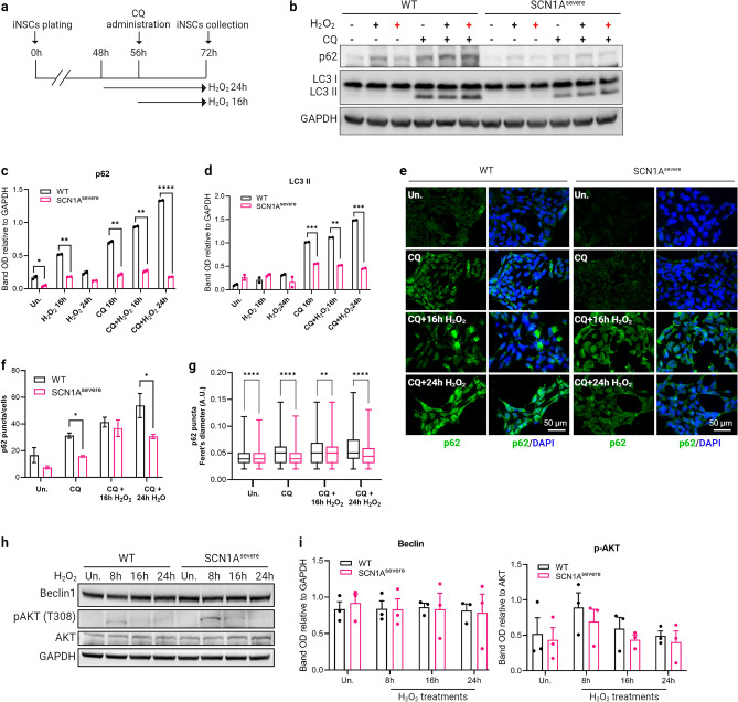
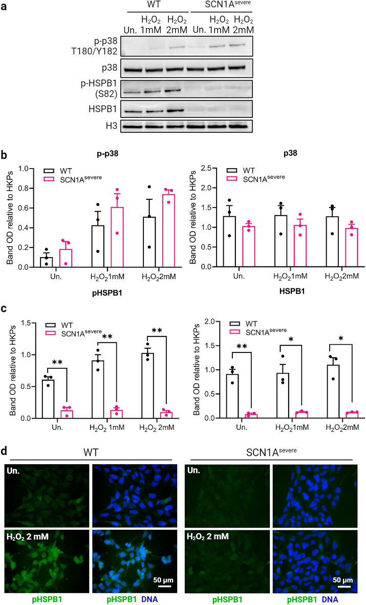
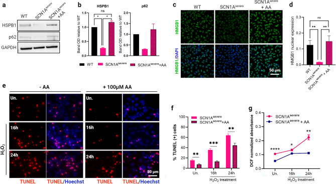
Prolonged febrile seizures (FS) in children are linked to the development of temporal lobe epilepsy (MTLE). The association between these two pathologies may be ascribed to the long-term effects that FS exert on neural stem cells, negatively affecting the generation of new neurons. Among the insults associated with FS, oxidative stress is noteworthy. Here, we investigated the consequences of exposure to hydrogen peroxide (H2O2) in an induced pluripotent stem cell-derived neural stem cells (iNSCs) model of a patient affected by FS and MTLE. In our study, we compare the findings from the MTLE patient with those derived from iNSCs of a sibling exhibiting a milder phenotype defined only by FS, as well as a healthy individual. In response to H2O2 treatment, iNSCs derived from MTLE patients demonstrated an elevated production of reactive oxygen species and increased apoptosis, despite the higher expression levels of antioxidant genes and proteins compared to other cell lines analysed. Among the potential causative mechanisms of enhanced vulnerability of MTLE patient iNSCs to oxidative stress, we found that these cells express low levels of the heat shock protein HSPB1 and of the autophagy adaptor SQSTM1/p62. Pre-treatment of diseased iNSCs with the antioxidant molecule ascorbic acid restored HSBP1 and p62 expression and simultaneously reduced the levels of ROS and apoptosis. Our findings suggest the potential for rescuing the impaired oxidative stress response in diseased iNSCs through antioxidant treatment, offering a promising mechanism to prevent FS degeneration in MTLE.


Conflict of interest statement

The authors declare no competing interests.

Figures

Figure 1
Figure 1
Effects of H2O2 treatment on iNSCs. (a) Schematic diagram showing experimental design for H2O2 treatment on iNSCs. (b) Healthy subject and epileptic patients iNSCs were treated with 2 mM H2O2 in a range of time from 30 min to 24 h. The production of ROS was assessed at each time point using DCFA-DA fluorescence, and a significantly higher level of ROS production in SCN1Asevere patient compared to healthy and SCN1Amild subjects was observed across all tested time intervals. DCFA-DA signal of each well was normalised to the number of cells in the same well. Data are presented as mean ± SEM of three biological replicates, *p < 0.05, **p < 0.01, ***p < 0.001, t-test has been calculated vs. WT in the same time point. (c) Analysis of expression of CAT gene in diseased and WT iNSCs under H2O2 treatment showed a significant increase only in SCN1Amild cells. (d) The expression of superoxide dismutase genes SOD1 and SOD3 was significantly up-regulated in SCN1Asevere patient iNSCs in respect to healthy and SCN1Amild cells at 8 and 16 h of H2O2 treatment, while (e) genes codifying for glutathione peroxidases GPX1 and GPX3 became significantly up-regulated in SCN1Asevere patient iNSCs in respect to healthy and SCN1Amild cells at 16 h of H2O2 treatments. For (ce), GAPDH was used as a housekeeping gene and data are represented as mean ± SEM of three biological replicates. *p < 0.05, **p < 0.01, ***p < 0.001, ****p < 0.0001, t-test. (f) Representative image of iNSCs stained with MitoTracker Green under H2O2 treatment of 8, 16 and 24 h. (g) The quantification of the size of mitochondria puncta showed a significant accumulation of these organelles under H2O2 treatment in SCN1Asevere iNSCs in respect to WT and SCN1Amild iNSCs. At least six images per condition were analysed. ***p < 0.001, t-test.
Figure 2
Figure 2
NRF2 expression is upregulated in SCN1Asevere patient iNSCs after H2O2 treatment. (a) The expression of NRF2 gene resulted significantly up-regulated in SCN1Asevere patient iNSCs in respect to WT and SCN1Amild cells at 16 h of 2 mM H2O2 treatments. (b) Immunoblot analysis showed that the NRF2 protein increased after 8, 16 and 24 h of H2O2 treatment in both WT and SCN1Asevere patient iNSCs, but for each time point tested we observed a higher expression of NRF2 in patient cells compared to WT cells. (c) Bands optical density (OD) of western blot showed in (b), calculated as NRF2 bands OD normalized on GAPDH band OD on the same time point. Data are presented as mean ± SEM of three biological replicates. (d) 8 and 16 h of H2O2 treatment induced a prominent NRF2 nuclear translocation in patient iNSCs in respect to SCN1Amild and healthy cells, as shown by quantification of NRF2 immunofluorescence signal overlapping with the nucleus (representative images are shown in Supplementary Fig. S2, at least five image per condition were analysed); *p < 0.05, **p < 0.01, ***p < 0.001, ****p < 0.0001, t-test. (e) Expression of NRF2 target genes NQO1 and HMOX1 after 8, 16 and 24 h of H2O2 treatment in WT and patients iNSCs. For qPCR, GAPDH was detected as loading control and data are presented as mean ± SEM of three biological replicates. *p < 0.05, **p < 0.01, ***p < 0.001, ****p < 0.0001, t-test.
Figure 3
Figure 3
SCN1Asevere iNSCs show increased apoptosis compared to SCN1Amild and healthy cells when treated with H2O2. (a) The apoptosis rate of iNSCs was measured using TUNEL assay after treatment with 2 mM H2O2 for 8, 16 and 24 h. The captured images showed apoptotic cells as bright red fluorescent cells. (b) Percentage of TUNEL positive cells calculated for each time point tested. TUNEL analysis showed that SCN1Asevere iNSCs treated with H2O2 displayed a significantly higher number of apoptotic cells compared to SCN1Amild and WT. At least 250 nuclei were analyzed for each condition.*p < 0.05, **p < 0.01, t-test. (c) After H2O2 treatment, the expression of pro-apoptotic gene BAX resulted significantly up-regulated in SCN1Asevere patient iNSCs compared to SCN1Amild and WT, while (d) the anti-apoptotic gene Bcl-2 exhibited an opposite trend, showing a down-regulation in SCN1Asevere iNSCs. For (c) and (d) panels, GAPDH was used as a housekeeping gene and *p < 0.05, **p < 0.01, ***p < 0.001, t-test. (e) Patient iNSCs exhibited an increased activation of caspase 3 by cleavage and reduced expression of the anti-apoptotic protein Bcl-xL after H2O2 treatments in all tested time points compared to WT cells. GAPDH was used as loading control. (fh) Quantification of protein band OD of western blot showed in (e), *p < 0.05, **p < 0.01, ***p < 0.001, t-test.
Figure 4
Figure 4
Oxidative stress-induced autophagy is impaired in SCN1Asevere patient iNSCs. (a) Schematic representation of H2O2 and CQ treatments on iNSCs. (b) Western blot analysis of protein involved in the autophagy process in cells treated with 2 mM H2O2 alone for 16 (black cross) and 24 h (red cross) or in combination with the autophagy inhibitor chloroquine (CQ). (c, d) Quantification of P62 and LC3II bands OD demonstrated that autophagy proteins accumulated in iNSCs of both SCN1Asevere patient and healthy subject when cells were simultaneously treated with H2O2 and CQ, but the expression of both proteins remained significantly lower in patient’s cells compared to WT in all time-point tested. *p < 0.05, **p < 0.01, ***p < 0.001, ****p < 0.0001, t-test. (e) The lowered expression of P62 protein in SCN1Asevere patient cells in respect to WT under H2O2 and CQ treatment was further confirmed by immunofluorescence analysis. (f) Quantification of p62 puncta and (g) of p62 puncta diameter in cells treated with H2O2 and CQ. (h) Western blot analysis of Beclin1 and of its inhibitor AKT under H2O2 treatment. (i) Quantification of western blot bands presented in (h) showed that there are no significant differences among the two cell lines in the activation of autophagy pathway under H2O2 treatment.
Figure 5
Figure 5
Down-regulation of HSBP1, a p38 downstream target, in SCN1Asevere iNSCs. (a) Western blot analysis of WT and SCN1Asevere iNSCs treated for 30 min with 1 or 2 mM H2O2 showed an activation of the stress-related kinase p38 by its phosphorylation at residues 180 and 182 in both cell lines tested. A higher activation of p38 in patient cells compared to WT was observed after H2O2 treatment. In the same blot, we detected the expression of the p38 target HSPB1. Interestingly, we found that this protein was expressed at very low levels in SCN1Asevere patient iNSCs compared to WT, in both total and phosphorylated form. GAPDH was used as loading control. (b, c) Quantification of biological replicates of western blot shown in (a). (d) pHSPB1 downregulation in SCN1Asevere cells under H2O2 treatments was further confirmed by immunofluorescence analysis.
Figure 6
Figure 6
Administration of ascorbic acid (AA) resulted in the restoration of HSPB1 and p62 levels and nuclear translocation of HMGB1, countering the effects of H2O2 in SCN1Asevere iNSCs. (a) The treatment of SCN1Asevere iNSCs with 100 μM AA for 12-h duration increase HSPB1 and p62 expression levels comparable to those observed in the WT cells. (b) Quantification of western blot bands shown in (a) presented as mean ± SEM of two biological replicates. *p < 0.05, t-test. (c, d) Treating SCN1Asevere cells with the antioxidant molecules AA led to nuclear translocation of HMGB1, a transcription factors interacting protein that regulates HSPB1 and p62 expression. **p < 0.01, t-test. (e) TUNEL assay in SCN1Asevere iNSCs treated with 2 mM H2O2 for 16 or 24 h showed a reduction of apoptotic cells (red fluorescence) in presence of AA pre-treatment (100 μM for 12 h) with respect to untreated condition. (f) Quantification of the number of TUNEL positive cells in experiment presented in (e); at least 300 nuclei were analysed for each condition. **p < 0.01, ***p < 0.001, t-test has been calculated vs. untreated condition in the same time point. (g) Production of ROS due to H2O2 treatment in diseased iNSCs in presence or absence of AA pre-treatment. AA significantly lowered levels of ROS across all tested time intervals. The DCFA-DA signal of each well was normalised to the number of cells in the same well. Data are presented as mean ± SEM of three biological replicates, *p < 0.05, **p < 0.01, ****p < 0.0001, t-test has been calculated vs. untreated in the same time point.

Similar articles

Cited by

References

    1. Anderson DJ. Stem cells and pattern formation in the nervous system: The possible versus the actual. Neuron. 2001;30:19–35. doi: 10.1016/S0896-6273(01)00260-4. - DOI - PubMed
    1. Kuhn H, Dickinson-Anson H, Gage F. Neurogenesis in the dentate gyrus of the adult rat: Age-related decrease of neuronal progenitor proliferation. J. Neurosci. 1996;16:2027–2033. doi: 10.1523/JNEUROSCI.16-06-02027.1996. - DOI - PMC - PubMed
    1. Huang T-T, Zou Y, Corniola R. Oxidative stress and adult neurogenesis—effects of radiation and superoxide dismutase deficiency. Semin. Cell Dev. Biol. 2012;23:738–744. doi: 10.1016/j.semcdb.2012.04.003. - DOI - PMC - PubMed
    1. Parent JM, Yu TW, Leibowitz RT, Geschwind DH, Sloviter RS, Lowenstein DH. Dentate granule cell neurogenesis is increased by seizures and contributes to aberrant network reorganization in the adult rat hippocampus. J. Neurosci. 1997;17:3727–3738. doi: 10.1523/JNEUROSCI.17-10-03727.1997. - DOI - PMC - PubMed
    1. Bengzon J, Kokaia Z, Elmér E, Nanobashvili A, Kokaia M, Lindvall O. Apoptosis and proliferation of dentate gyrus neurons after single and intermittent limbic seizures. Proc. Natl. Acad. Sci. USA. 1997;94:10432–10437. doi: 10.1073/pnas.94.19.10432. - DOI - PMC - PubMed

MeSH terms