. 2024 Mar 12;81(1):125.
doi: 10.1007/s00018-024-05167-6.

Vascular ATGL-dependent lipolysis and the activation of cPLA2-PGI2 pathway protect against postprandial endothelial dysfunction

Affiliations

Vascular ATGL-dependent lipolysis and the activation of cPLA2-PGI2 pathway protect against postprandial endothelial dysfunction

M Sternak et al. Cell Mol Life Sci. .

Abstract

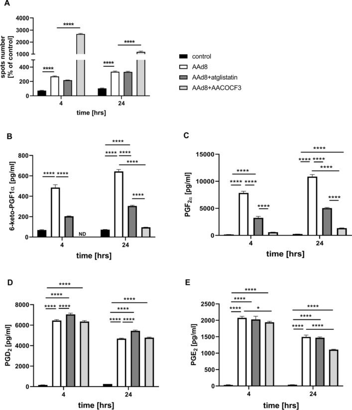
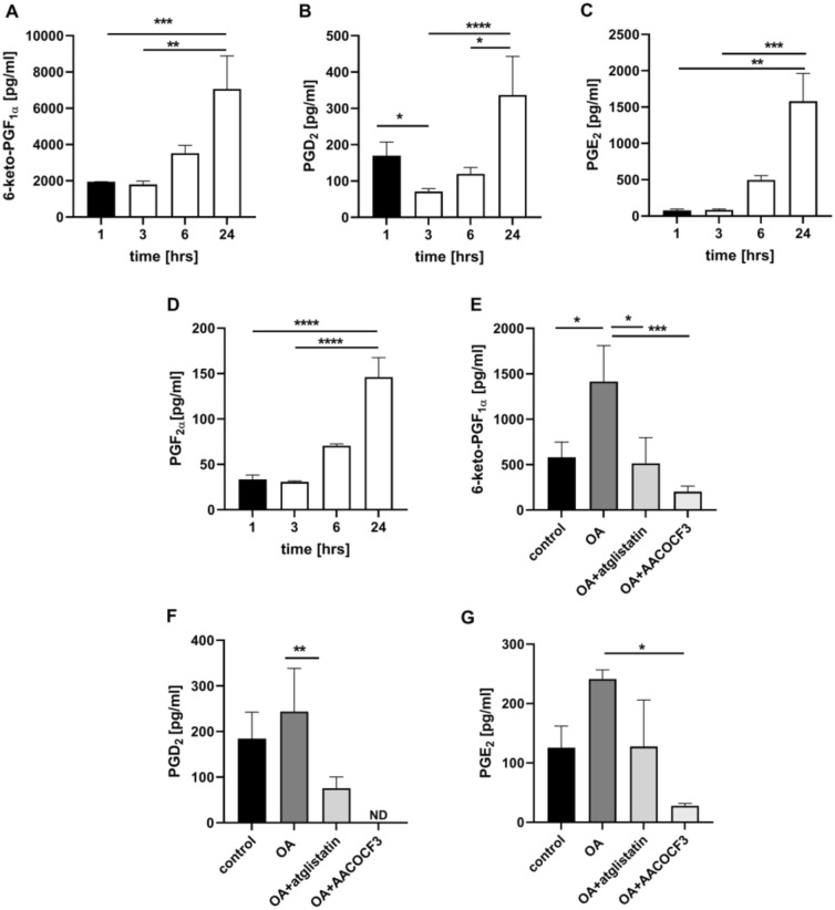
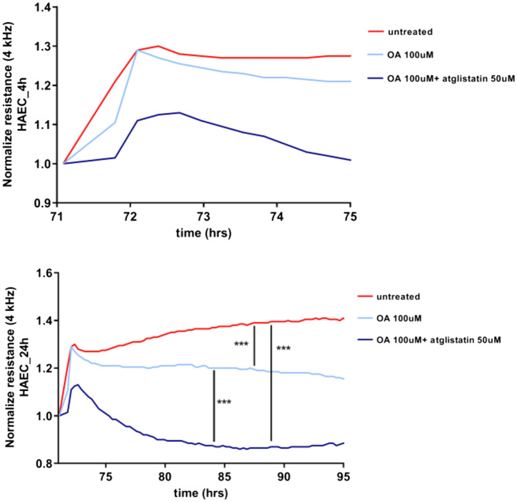
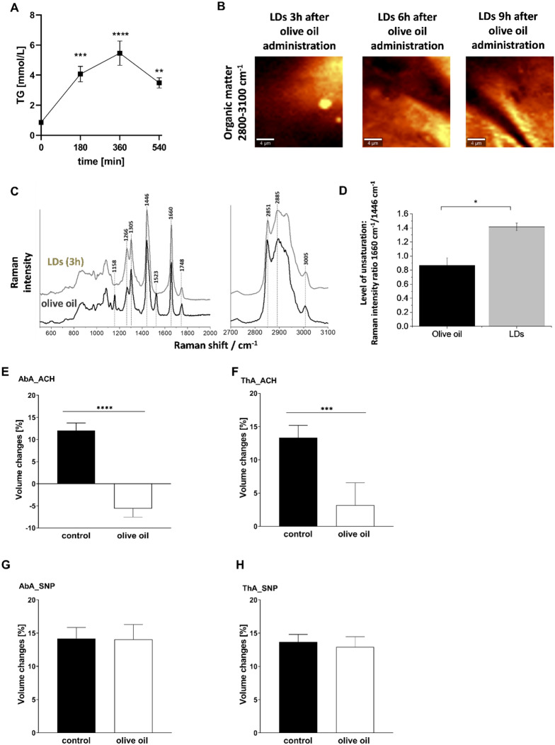
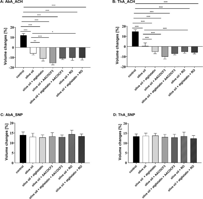
Adipose triglyceride lipase (ATGL) is involved in lipolysis and displays a detrimental pathophysiological role in cardio-metabolic diseases. However, the organo-protective effects of ATGL-induced lipolysis were also suggested. The aim of this work was to characterize the function of lipid droplets (LDs) and ATGL-induced lipolysis in the regulation of endothelial function. ATGL-dependent LDs hydrolysis and cytosolic phospholipase A2 (cPLA2)-derived eicosanoids production were studied in the aorta, endothelial and smooth muscle cells exposed to exogenous oleic acid (OA) or arachidonic acid (AA). Functional effects of ATGL-dependent lipolysis and subsequent activation of cPLA2/PGI2 pathway were also studied in vivo in relation to postprandial endothelial dysfunction.The formation of LDs was invariably associated with elevated production of endogenous AA-derived prostacyclin (PGI2). In the presence of the inhibitor of ATGL or the inhibitor of cytosolic phospholipase A2, the production of eicosanoids was reduced, with a concomitant increase in the number of LDs. OA administration impaired endothelial barrier integrity in vitro that was further impaired if OA was given together with ATGL inhibitor. Importantly, in vivo, olive oil induced postprandial endothelial dysfunction that was significantly deteriorated by ATGL inhibition, cPLA2 inhibition or by prostacyclin (IP) receptor blockade.In summary, vascular LDs formation induced by exogenous AA or OA was associated with ATGL- and cPLA2-dependent PGI2 production from endogenous AA. The inhibition of ATGL resulted in an impairment of endothelial barrier function in vitro. The inhibition of ATGL-cPLA2-PGI2 dependent pathway resulted in the deterioration of endothelial function upon exposure to olive oil in vivo. In conclusion, vascular ATGL-cPLA2-PGI2 dependent pathway activated by lipid overload and linked to LDs formation in endothelium and smooth muscle cells has a vasoprotective role by counterbalancing detrimental effects of lipid overload on endothelial function.

Keywords: ATGL; Atglistatin; Endothelial-induced vasodilation; Endothelium; Lipid droplets; Lipolysis.


Conflict of interest statement

Not applicable.

Figures

Fig. 1
Fig. 1
Lipid droplets formation in human aortic endothelial cells (HAEC) induced by exogenous AA (AAd8) and ATGL- and cPLA2-dependent eicosanoid release from endogenous AA. Effect of inhibition of atglistatin and AACOCF3 on lipid droplets formation (A) and eicosanoids release (BE) in HAEC  4 h and 24 h after deuterated arachidonic acid (AAd8, 25 µM) addition in the presence or absence of atglistatin (50 µM) and AACOCF3 (10 µM). Data represent mean ± SD of three independent experiments. Statistical analysis was performed using one-way ANOVA followed by Tukey’s multiple comparisons test (*p < 0.05, ****p < 0.0001). ND - not detected
Fig. 2
Fig. 2
The content of egxogenous AAd8 and endogenous AA in lipid extracts and synthesis of PGI2 from endogenous AA by cPLA2/COX-1/COX-2 pathway in response to exogenous AAd8 in human aortic endothelial cells (HAEC). The content of exogenous deuterated arachidonic acid (AAd8, 25 µM) (A), endogenous AA (B) in lipid extracts and the concentration of 6-keto-PGF (C) in medium in the presence or absence of cPLA2, COX-1, COX-2 inhibitors (AACOCF3, SC-560, DuP-697) after 4 h and 24 h of HAEC incubation with AAd8. Data represent mean ± SD of three independent experiments. Statistical analysis was performed using the nonparametric Kruskal–Wallis test followed by Dunn’s multiple comparisons test (*p < 0.05, **p < 0.01, ***p < 0.001, ****p < 0.0001). ND - not detected
Fig. 3
Fig. 3
Lipid droplets formation in human aortic endothelial cells (HAEC) and vascular smooth muscle cells (MOVAS) induced by exogenous OA; parallel formation of PGI2 from endogenous AA in vitro.Time course of changes in lipid droplets formation (A, B) and 6-keto-PGF release (C, D) stimulated by deuterated OA (OAd34, 100M, 1 h, 2 h, 3 h, 6 h, 24 h). Data represent mean ± SD of three independent experiments. Statistical analysis was performed using one-way ANOVA followed by Tukey’s or Bonferroni’s multiple comparisons test (*p < 0.05, **p < 0.01, ****p < 0.0001)
Fig. 4
Fig. 4
Lipid droplets formation in isolated murine aorta in response to exogenous OA. Time course of changes in lipid droplets formation in endothelium (A) and smooth muscle cells (B) within isolated murine aorta stimulated by deuterated oleic acid (OAd34, 1 mM; 1 h, 2 h, 3 h, 6 h, 24 h). The data are presented as the median with interquartile range (n = 4). Statistics: Kruskal–Wallis test followed by Dunn’s multiple comparisons test. (*p< 0.05, **p < 0.01 ). Effect of inhibition of atglistatin and AACOCF3 on lipid droplets formation in isolated murine aorta ex vivo stimulated by OA (500 µM, 24 h) in the presence or absence of atglistatin (50 µM) and AACOCF3 (10 µM)(C). The data are presented as the median with interquartile range (n = 6–8). Statistics: Kruskal–Wallis test followed by Dunn’s multiple comparisons test (*p < 0.05, **p < 0.01, ***p < 0.001). Representative microphotographs of immunostaining of control en face aorta and aorta treated with OA (1 mM, 24 h). Red, blue and green fluorescence originating from PECAM-1, cell nuclei and LDs, respectively (D). Visualisation of biochemical components of murine en face aorta with LDs formed in response to OAd34 (1 mM, 24 h) by Raman spectroscopy (brighter colours characterizing higher intensities of respective bands). Apart from typical structures like nuclei or elastin fibres Raman imaging of en face aorta show two kinds of lipid droplets: rich in OAd34 and without OAd34 (E, F) but most likely containing AA
Fig. 5
Fig. 5
ATGL- and cPLA2-dependent PGI2 release from endogenous AA in isolated murine aorta in response to exogenous OA. Time course of changes in eicosanoids release in endothelium within isolated murine aorta stimulated by deuterated oleic acid (OAd34, 1 mM; 1 h, 2 h, 3 h, 6 h, 24 h) (AD). The data are presented as the median with interquartile range (n = 6). Statistics: Kruskal–Wallis test followed by Dunn’s multiple comparisons test (*p< 0.05, **p < 0.01, ***p < 0.001, ****p < 0.0001). Effect of inhibition of atglistatin (50µM) and AACOCF3 (10 µM) on eicosanoids release in isolated murine aorta ex vivo stimulated by OA (500 µM, 24 h) (EG). The results are presented as the mean ± SD (n = 6–14). Statistics: one-way ANOVA followed by Tukey’s or Bonferroni’s multiple comparisons test) (*p < 0.05, **p < 0.01, ***p < 0.001, ****p < 0.0001)
Fig. 6
Fig. 6
Effects of the inhibition of ATGL-dependent-lipolysis of LDs induced by exogenous OA on the regulation of the endothelial barrier integrity in vitro. Effect of inhibition of atglistatin (50 µM) on permeabililty changes in HAEC 4 h and 24 h after OA administration (100 µM) measured by using ECIS method. Data represent mean ± SD of three independent experiments Statistics: one-way ANOVA followed by Tukey’s multiple comparisons test (***p < 0.001)
Fig. 7
Fig. 7
Postprandial formation of LD and development of endothelial dysfunction in vivo in aorta associated with increased TG plasma level induced by olive oil gavage. C57BL/6 mice were fasted for 16 h before olive oil administration. The dose of olive oil was chosen based on plasma TG measurement with plasma TG levels peak at 360 min (A). The results are presented as the mean ± SD (n = 4–6). Statistics: one-way ANOVA followed by Tukey’s multiple comparisons test (**p < 0.01, ***p < 0.001, ****p < 0.0001). Raman imaging of lipid droplets formation in murine aorta isolated from mice 3 h, 6 h and 9 h after intra-gastric administration of the olive oil (10 mL/mg) (B). Comparison of the spectrum of the olive oil with the average spectrum of endothelial LDs of an isolated blood vessel 3 h, after intra-gastric administration of the olive oil. The Raman spectrum of LDs indicates higher lipid unsaturation than the administered olive oil as the ratio of the bands 1660 to 1446 cm−1, and 1266 to 1305 cm−1 is higher for the LDs spectrum than for olive oil (C, D). Endothelial function after olive oil administration—assessed in vivo using unique MRI-based analysis (E). Changes in end-diastolic volume of the abdominal (A:AbA) and thoracic aorta (B: ThA) 30 min after acetylcholine administration or 30 min after sodium nitroprusside administration (C: AbA; D: ThA) in C57BL/6 mice gavaged with olive oil (10 mL/kg). Measurements were placed 6 h after the olive oil gavave. The dose of olive oil was chosen based on the plasma triglycerides measurement curve. The results are presented as the mean ± SD (n = 6–14). Statistics: one-way ANOVA followed by Tukey’s multiple comparisons test (*p < 0.05, ***p < 0.001)
Fig. 8
Fig. 8
Protective role of vascular ATGL-dependent lipolysis and the activation of cPLA2–PGI2 pathway in the postprandial endothelial dysfunction induced by olive oil gavage. Atglistatin (200 µg/mL), AACOCF3 (10 mg/kg) and RO 3244794 (50 mg/kg) were used to inhibit ATGL, cPLA2 or to block IP receptor respectively. Endothelial function after olive oil administration was assessed in vivo using unique MRI-based analysis (AC). Changes in end-diastolic volume of the abdominal (A: AbA) and thoracic aorta (B: ThA) were measured 30 min after acetylcholine administration or 30 min after sodium nitroprusside administration (C: AbA; D: ThA) in C57BL/6 mice gavaged with olive oil (10 mL/kg). Measurements were performed 6 h after olive oil gavage. Statistics: one-way ANOVA followed by Tukey’s multiple comparisons test (*p < 0.05, **p < 0.01, ***p < 0.001)

Similar articles

Cited by

References

    1. Schreiber R, Xie H, Schweiger M. Of mice and men: the physiological role of adipose triglyceride lipase (ATGL) Biochim Biophys Acta Mol Cell Biol Lipids. 2019;1864:880–899. doi: 10.1016/j.bbalip.2018.10.008. - DOI - PMC - PubMed
    1. Schweiger M, Romauch M, Schreiber R, Grabner GF, Hütter S, Kotzbeck P, Benedikt P, Eichmann TO, Yamada S, Knittelfelder O, Diwoky C, Doler C, Mayer N, De Cecco W, Breinbauer R, Zimmermann R, Zechner R. Pharmacological inhibition of adipose triglyceride lipase corrects high-fat diet-induced insulin resistance and hepatosteatosis in mice. Nat Commun. 2017;8:14859. doi: 10.1038/ncomms14859. - DOI - PMC - PubMed
    1. Kaur S, Auger C, Barayan D, Shah P, Matveev A, Knuth CM, Harris TE, Jeschke MG. Adipose-specific ATGL ablation reduces burn injury-induced metabolic derangements in mice. Clin Transl Med. 2021;11:417. doi: 10.1002/ctm2.417. - DOI - PMC - PubMed
    1. Bottermann K, Granade ME, Oenarto V, Fischer JW, Harris TE. Atglistatin pretreatment preserves remote myocardium function following myocardial infarction. J Cardiovasc Pharmacol Ther. 2021;26:289–297. doi: 10.1177/1074248420971113. - DOI - PubMed
    1. Thiele A, Luettges K, Ritter D, Beyhoff N, Smeir E, Grune J, Steinhoff JS, Schupp M, Klopfleisch R, Rothe M, Wilck N, Bartolomaeus H, Migglautsch AK, Breinbauer R, Kershaw EE, Grabner GF, Zechner R, Kintscher U, Foryst-Ludwig A. Pharmacological inhibition of adipose tissue adipose triglyceride lipase by Atglistatin prevents catecholamine-induced myocardial damage. Cardiovasc Res. 2021;118:2488. doi: 10.1093/cvr/cvab182. - DOI - PMC - PubMed

LinkOut - more resources