

Validation of anterior ankle soft tissue dynamics and shear modulus for anterior ankle impingement syndrome after ankle fracture surgery
- PMID: 38467787
- PMCID: PMC10928075
- DOI: 10.1038/s41598-024-56671-5
Validation of anterior ankle soft tissue dynamics and shear modulus for anterior ankle impingement syndrome after ankle fracture surgery
Abstract
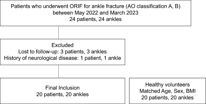
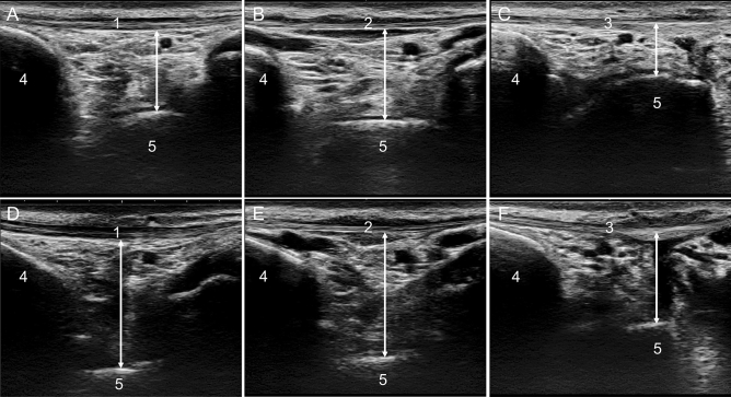
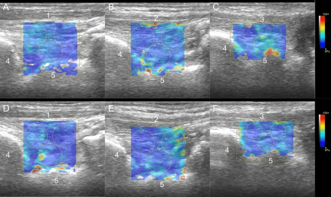
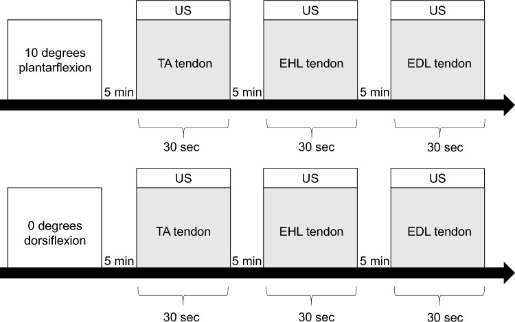
Anterior ankle impingement syndrome (AAIS) has been reported to account for a high percentage of complications following ankle fracture surgery. The soft tissue etiology of AAIS is thought to be thickening and inflammation of the anterior ankle soft tissues intervening anteriorly at the tibiotalar joint, causing pain and functional limitation during dorsiflexion. However, the effects of anterior ankle soft tissue dynamics and stiffness on AAIS have yet to be clarified. This study aimed to determine the relationship between AAIS and the anterior ankle soft tissue thickness change ratio and shear modulus using ultrasonography (US). The participants were 20 patients with ankle joint fractures (AO classification A, B) who had undergone open reduction and internal fixation and 20 healthy adults. The evaluation periods were 3 months and 6 months postoperatively. US was used to delineate the tibialis anterior tendon, extensor hallucis longus tendon, and the extensor digitorum longus tendon over the talus and tibia on a long-axis image. Anterior ankle soft tissue thickness was measured as the shortest distance from the most convex part of the talus to the tendon directly above it. The Anterior ankle soft tissue thickness change ratio was determined by dividing the value at 0° dorsiflexion by the value at 10° plantarflexion. The same images as for the anterior soft tissue thickness measurement were drawn for the shear modulus measurement, and the average shear modulus (kPa) was calculated using shear-wave elastography. There was no significant difference in the thickness change ratio between the postoperative and healthy groups. Compared with the healthy group, the shear modulus was significantly higher at 3 and 6 months in the postoperative group (p < 0.01). The shear elastic modulus at 6-month postoperative group was significantly lower than at 3-month postoperative group (p < 0.01). Anterior ankle joint soft tissue stiffness may increase after surgery for an ankle fracture.
Keywords: Ankle fracture; Anterior ankle impingement syndrome; Anterior ankle soft tissue; Shear-wave elastography; Ultrasonography.
© 2024. The Author(s).
Conflict of interest statement
The authors declare no competing interests.
Figures
Similar articles
-
The reliability of shear elastic modulus measurement of the ankle plantar flexion muscles is higher at dorsiflexed position of the ankle.J Foot Ankle Res. 2017 Apr 18;10:18. doi: 10.1186/s13047-017-0199-0. eCollection 2017. J Foot Ankle Res. 2017. PMID: 28428826 Free PMC article.
-
[The Postero-Lateral Approach--An Alternative to Closed Anterior-Posterior Screw Fixation of a Dislocated Postero-Lateral Fragment of the Distal Tibia in Complex Ankle Fractures].Z Orthop Unfall. 2015 Jun;153(3):289-95. doi: 10.1055/s-0035-1545706. Epub 2015 May 6. Z Orthop Unfall. 2015. PMID: 25959570 Clinical Trial. German.
-
Etiology and Treatment of Delayed-Onset Medial Malleolar Pain Following Total Ankle Arthroplasty.Foot Ankle Int. 2016 Aug;37(8):822-8. doi: 10.1177/1071100716643278. Epub 2016 Apr 6. Foot Ankle Int. 2016. PMID: 27053405
-
The distal fascicle of the anterior inferior tibiofibular ligament as a cause of tibiotalar impingement syndrome: a current concepts review.Knee Surg Sports Traumatol Arthrosc. 2007 Apr;15(4):465-71. doi: 10.1007/s00167-006-0275-7. Epub 2007 Jan 20. Knee Surg Sports Traumatol Arthrosc. 2007. PMID: 17237964 Free PMC article. Review.
-
Ankle impingement: a review of multimodality imaging approach.Musculoskelet Surg. 2013 Aug;97 Suppl 2:S161-8. doi: 10.1007/s12306-013-0286-8. Epub 2013 Aug 15. Musculoskelet Surg. 2013. PMID: 23949938 Review.
References
MeSH terms
Grants and funding
LinkOut - more resources
Full Text Sources
Medical
