Review
. 2024 Apr;16(4):700-714.
doi: 10.1038/s44321-024-00045-x. Epub 2024 Mar 11.

The 'communicatome' of pregnancy: spotlight on cellular and extravesicular chimerism

Affiliations
Review

The 'communicatome' of pregnancy: spotlight on cellular and extravesicular chimerism

Isabel Graf et al. EMBO Mol Med. 2024 Apr.

Abstract

Communication via biological mediators between mother and fetus are key to reproductive success and offspring's future health. The repertoire of mediators coding signals between mother and fetus is broad and includes soluble factors, membrane-bound particles and immune as well as non-immune cells. Based on the emergence of technological advancements over the last years, considerable progress has been made toward deciphering the "communicatome" between fetus and mother during pregnancy and even after birth. In this context, pregnancy-associated chimerism has sparked the attention among immunologists, since chimeric cells-although low in number-are maintained in the allogeneic host (mother or fetus) for years after birth. Other non-cellular structures of chimerism, e.g. extracellular vesicles (EVs), are increasingly recognized as modulators of pregnancy outcome and offspring's health. We here discuss the origin, distribution and function of pregnancy-acquired microchimerism and chimeric EVs in mother and offspring. We also highlight the pioneering concept of maternal microchimeric cell-derived EVs in offspring. Such insights expand the understanding of pregnancy-associated health or disease risks in mother and offspring.

Keywords: Extracellular Vesicles; Feto-maternal Communication; Microchimerism; Pregnancy.


Conflict of interest statement

The authors declare no competing interests.

Figures
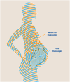
Figure 1
Figure 1. The “communicatome” of pregnancy.
Maternal (marked in orange) as well as fetal (blue) mediators, e.g., antibodies, hormones, cells, extracellular vesicles, are vertically transferred during pregnancy. Due to the longevity of some of these mediators, e.g., cells, this feto-maternal communicatome can have long-lasting impacts on maternal and offspring’s health.
Figure 2
Figure 2. The chimeric coding systems—features and functions.
Characteristics of the pregnancy-acquired chimeric coding systems, focusing on extracellular vesicles and cells (left) and their associated characteristics, features and functions (right).
Figure 3
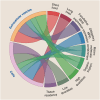
Figure 3. Implications of maternal microchimeric cells and chimeric extracellular vesicles.
Pregnancy-associated (micro-)chimerism is associated with numerous consequences for fetal health. The inner circle depicts mechanisms by which corresponding outcomes—displayed in the outer circle—are promoted by either EVs (small dots), microchimeric cells (large dots), or both. For example, there is a MMc-associated modulation of hematopoetic differentiation, which protects the infant against infections.
Figure 4
Figure 4. Implications of fetal microchimeric cells and chimeric extracellular vesicles.
Pregnancy-associated (micro-)chimerism is associated with numerous consequences for maternal health. The inner circle depicts mechanisms by which corresponding outcomes—displayed in the outer circle—are promoted by either EVs (small dots), microchimeric cells (large dots), or both. For example, an EV-associated immune dysregulation is connected to the modulation of pregnancy outcomes as well as autoimmune diseases.
Figure 5
Figure 5. Extracellular vesicles—biogenesis and cargo.
There are four major subtypes of vesicles released into the extracellular space when referring to placenta-associated EVs: exosomes, ectosomes, apoptotic bodies and syncytial nuclear aggregates (left part of this figure). Exosomes are generated via the endosomal pathway (left top). Starting point is the production of early endosomes by invagination of the cell plasma membrane containing plasma membrane proteins as well as extracellular proteins and eventually they mature to late endosomes (Mathieu et al, 2019). The endosomes are then loaded with proteins originating from the trans golgi network as well as the endoplasmatic reticulum and vice versa release proteins to these compartments. Next, by inward budding of the membrane of the late endosomes multi vesicular bodies (MVB) arise, which form the EV precursors. Fusion of MVB with the plasma membrane finally leads to release of EVs termed as exosomes, which are in the size range of around 50–150 nm. The second major EV biogenesis pathway consists of outward budding of the plasma membrane with consecutive shedding of the vesicle (right top). Also here cargo sorting mechanisms direct the future cargo of the EVs to the plasma membrane (Tricarico et al, 2017). These EVs are termed ectosomes with the microvesicles being their best studied representative and range from 200 nm up to >1000 nm. Among the exosomes and ectosomes a considerable number of subpopulations has been identified: large oncosomes, exophers, mirgasomes—just to mention a few (Buzas, 2023). The apoptosis of a cell leads to the third major EV population: apoptotic bodies (right buttom). They are rather variable in content and size and range from 100–5000 nm. Lastly, the outer layer of the placenta—the syncytiotrophoblast, which can be characterized by its multinuclear syncytium—secretes vesicles larger >20 µm containing several nuclei (left bottom) (Huppertz et al, ; Tong et al, 2018). The cargo of EVs includes proteins, lipids, DNA and RNA (right part of this figure). The cargo varies depending on the specific EV subtype. In addition, it has been suggested that EVs are surrounded by a protein corona referring to proteins and other macromolecules, which are externally attached to the EV and form a surrounding layer (Heidarzadeh et al, 2023). These macromolecules might spontaneously adsorb to the EVs’ surface and are acquired from protein-rich environments such as plasma (Tóth et al, 2021). PLAP placental alkaline phosphatase, sEVs small EVs, lEVs large EVs.
Figure 6
Figure 6. Induction of split tolerance by the presentation of MMc-derived EV.
EVs released by MMc carry MHC molecules (red receptor), which can be taken up by antigen-presenting cells, where they are either incorporated into the plasma membrane (left) or processed and presented as antigen (right). The process of MHC molecule incorporation is referred to as cross-dressing and simultaneously leads to an upregulation and co-localization of CD86—an activator of proliferation (Bracamonte-Baran et al, 2017). This results in an activation of T cells upon T-cell receptor-specific (blue receptor) presentation of peptides (orange). Processing and presentation of the antigen (right) was associated with an emergence and co-localization of PD-L1—an inhibitor of cell proliferation (Bracamonte-Baran et al, 2017). As a result, interaction with T cells and their respective T-cell receptors (blue receptor) specific for these allopeptides (red peptide) led to an abortive activation. The selective upregulation and localization of co-stimulatory molecules depending on the MHC being cross-dressed or presented as allopeptide, leads to a divergent “split immune reaction”.

Similar articles

Cited by

References

    1. Albrecht M, Pagenkemper M, Wiessner C, Spohn M, Lütgehetmann M, Jacobsen H, Gabriel G, Zazara DE, Haertel C, Hecher K, et al. Infant immunity against viral infections is advanced by the placenta-dependent vertical transfer of maternal antibodies. Vaccine. 2022;40:1563–1571. doi: 10.1016/j.vaccine.2020.12.049. - DOI - PubMed
    1. Amenta PS, Gay S, Vaheri A, Martinez-Hernandez A. The extracellular matrix is an integrated unit: ultrastructural localization of collagen types I, III, IV, V, VI, fibronectin, and laminin in human term placenta. Coll Relat Res. 1986;6:125–152. doi: 10.1016/S0174-173X(86)80021-8. - DOI - PubMed
    1. Ariga H, Ohto H, Busch MP, Imamura S, Watson R, Reed W, Lee TH. Kinetics of fetal cellular and cell-free DNA in the maternal circulation during and after pregnancy: implications for noninvasive prenatal diagnosis. Transfusion. 2001;41:1524–1530. doi: 10.1046/j.1537-2995.2001.41121524.x. - DOI - PubMed
    1. Arutyunyan A, Roberts K, Troulé K, Wong FCK, Sheridan MA, Kats I, Garcia-Alonso L, Velten B, Hoo R, Ruiz-Morales ER, et al. Spatial multiomics map of trophoblast development in early pregnancy. Nature. 2023;616:143–151. doi: 10.1038/s41586-023-05869-0. - DOI - PMC - PubMed
    1. Auber M, Svenningsen P. An estimate of extracellular vesicle secretion rates of human blood cells. J Extracell Biol. 2022;1:e46. doi: 10.1002/jex2.46. - DOI - PMC - PubMed