Review
. 2024 Jun;21(6):428-443.
doi: 10.1038/s41575-024-00906-3. Epub 2024 Mar 11.

Group 3 innate lymphoid cells in intestinal health and disease

Affiliations
Review

Group 3 innate lymphoid cells in intestinal health and disease

Veronika Horn et al. Nat Rev Gastroenterol Hepatol. 2024 Jun.

Abstract

The gastrointestinal tract is an immunologically rich organ, containing complex cell networks and dense lymphoid structures that safeguard this large absorptive barrier from pathogens, contribute to tissue physiology and support mucosal healing. Simultaneously, the immune system must remain tolerant to innocuous dietary antigens and trillions of normally beneficial microorganisms colonizing the intestine. Indeed, a dysfunctional immune response in the intestine underlies the pathogenesis of numerous local and systemic diseases, including inflammatory bowel disease, food allergy, chronic enteric infections or cancers. Here, we discuss group 3 innate lymphoid cells (ILC3s), which have emerged as orchestrators of tissue physiology, immunity, inflammation, tolerance and malignancy in the gastrointestinal tract. ILC3s are abundant in the developing and healthy intestine but their numbers or function are altered during chronic disease and cancer. The latest studies provide new insights into the mechanisms by which ILC3s fundamentally shape intestinal homeostasis or disease pathophysiology, and often this functional dichotomy depends on context and complex interactions with other cell types or microorganisms. Finally, we consider how this knowledge could be harnessed to improve current treatments or provoke new opportunities for therapeutic intervention to promote gut health.


Figures

Figure 1 ∣
Figure 1 ∣. Phenotype and spatiotemporal properties of ILC3 subsets in the human and mouse intestine.
a. Mouse group 3 innate lymphoid cells (ILC3s) are RORγt+ CD127+ c-kit+ and are further distinguished by expression of surface markers, transcription factors, signature cytokines, and anatomical distribution in the gut. LTi-like ILC3s accumulate in lymphoid clusters termed cryptopatches prior to birth, highly express CCR6, are heterogeneous in CD4, and lack NKp46 or T-bet. cILC3s are scattered throughout the lamina propria and often co-express NKp46 and T-bet. Upon inflammatory stimuli, cILC3s can transdifferentiate into ex-ILC3s that lose RORγt but maintain T-bet and produce high amounts of IFNγ. Additionally, inflammatory ILC3s (iILC3) that co-express RORγt and T-bet, but not CD4 or NKp46, have been described in colitis, neuroinflammation, and myocarditis, and are likely derived from the circulation. b. Human ILC3s are RORγt+ CD127+ CD161+ c-kit+. Much less is known about the heterogeneity of human ILC3s, for which NKp44 seems to be a marker of activation. Human ILC3s can differentiate into a ILC1-like ex-ILC3 phenotype. NKp44 ILC3s likely contain Nrp1+ LTi-like ILC3s, but additional research is needed to determine this subset in humans. c. LTi and LTi-like ILCs are abundant before birth, and in mice, peak around weaning and decline steadily across life. cILC3s develop postnatally in response to microbiota and nutrients and become the dominant subset with advanced age. Across life, adaptive effector cells become more abundant in the intestine. Human ILC3s decline with age and chronic infectious or inflammatory challenges, but subset distribution remains unclear, and is therefore not shown here. GM-CSF, granulocyte–macrophage colony-stimulating factor; HB-EGF, heparin-binding EGF-like growth factor; LTα1β2; lymphotoxin α1β2; MHCII, major histocompatibility complex class II; NRP1, neuropilin 1.
Figure 2 ∣
Figure 2 ∣. ILC3s orchestrate tissue physiology and immune tolerance in the gut.
Intestinal group 3 innate lymphoid cells (ILC3s) interact with several immune and non-immune cell modules to promote intestinal barrier integrity, anti-microbial responses, immune tolerance, or inflammation. For example, ILC3s produce IL-22, IL-17, and IFNγ, which act on intestinal epithelial cells (IECs) to produce antimicrobial peptides such as as RegIIIγ, S100A, and lipocalin, and mucus to support bacterial clearance or shape gut microbiota composition. ILC3s are regulated by luminal nutrients such as retinoic acid or vitamin D and microbial metabolites such as short-chain fatty acids (SCFAs). Additionally, ILC3s react to innate-derived cytokines IL-1, IL-23, and TL1A, produced by CX3CR1+ mononuclear phagocytes (MNP), and in turn produce GM-CSF to regulate MNP responses. ILC3s further support a balance between tolerogenic regulatory T (Treg) cells and pathogenic type 17 T helper (Th17) cells via MHCII-dependent antigen presentation and IL-2 production to promote tolerance towards dietary antigens and microbiota. Neuron-derived factors such as acetylcholine, noradrenaline, vasoactive intestinal peptide (VIP), and glial derived neurotrophic factor (GDNF) family ligands further fine-tune ILC3 responses. Finally, ILC3s are dependent on a mesenchymal stroma cell nice that provides IL-7 and thymic stromal lymphopoietin (TLSP), which in turn relies on surface bound lymphotoxin-β (LTβ) on LTi-like ILC3.
Figure 3 ∣
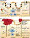
Figure 3 ∣. ILC3s in human IBD and CRC.
a. In inflammatory bowel disease (IBD), a breakdown of the intestinal barrier and increased intestinal epithelial cell (IEC) death leads to TNF-mediated activation of mononuclear phagocytes (MNPs), which produce IL-1, IL-23, and TL1A. In this highly proinflammatory milieu, ILC3s become depleted or dysfunctional by unknown mechanisms, likely involving plasticity, cell death, or subset re-distribution. The pro-inflammatory environment increases ILC3 plasticity towards a pathogenic ex-ILC3 phenotype, and accumulation of inflammatory ILC3s (iILC3s), which produce proinflammatory cytokines and sustain inflammation. ILC3 depletion and dysfunction further supports dysregulated adaptive responses which further increase inflammation. Conversely, ILC3s or related pathways are targeted by all current or upcoming biologic therapies including anti-TNF, anti-IL23(p19), IL-12/23(p40), or TL1A, as highlighted by white boxes. ILC3s also express, and potentially gut home via α4β7-integrin, which could be affected by anti-integrin treatment, and sphingosine-1-phosphate receptor (S1PR), which is targeted by anti-S1PR biologics. Furthermore, cytokine production by ILC3s, such as IL-22, depends on JAK–STAT signaling which could be affected by JAK/STAT small molecule inhibitors. b. In colorectal cancer (CRC), ILC3s promote various antitumor effects in early stage of tumorigenesis via key effector cytokines. This step includes IL-22 mediated stem cell protection from genotoxic stress, intestinal repair, and lymphotoxin-mediated tertiary lymphoid structure (TLS) formation. Conversely, the inflammatory tumor microenvironment leads to a depletion of ILC3s and the remaining cells show dysfunctional features, exhaustion, and increased plasticity towards an ILC1-like, ex-ILC3 phenotype. The role of ILC3-derived IL-22 in CRC is dichotomous, since production of IL-22 can limit or promote tumor progression depending on timepoint of tumorigenesis and availability of its antagonist IL-22BP. Impaired ILC3s results in compositional shifts in the microbiota, and this reduces type-1 immunity in CD4+ and CD8+ T cells, which enhances tumor progression or invasion, as well as causes resistance to anti-PD-1-directed therapies. Moreover, ILC3s express PD1, and could be targeted by anti-PD1 therapies or contribute to checkpoint-associated colitis but this has not yet been extensively explored. HB-EGF, heparin-binding EGF-like growth factor; cILC3s, conventional ILC3s; S1PR, S1P receptor.
Figure 4 ∣
Figure 4 ∣. Harnessing ILC3s as a novel therapy to promote gut health.
Although in early stages of investigation, it remains possible to boost tolerogenic group 3 innate lymphoid cells (ILC3) subsets to promote gut health. This approach could be accomplished by fecal microbiota transplantation (FMT), probiotics, specifically enhancing protective ILC3 responses with small molecules, or through a cell-based transfer approach. Functional ILC3s could further resolve the imbalance between proinflammatory Th17 and tolerogenic Treg cells by educating appropriate adaptive responses. Ultimately, ILC3s can promote restoration of the intestinal barrier and mucosal healing by production of IL-22 and HB-EGF.

References

    1. Spits H. et al. Innate lymphoid cells--a proposal for uniform nomenclature. Nat. Rev. Immunol 13, 145–149 (2013). - PubMed
    1. Sonnenberg GF & Artis D Innate lymphoid cells in the initiation, regulation and resolution of inflammation. Nat. Med 21, 698–708 (2015). - PMC - PubMed
    1. Vivier E. et al. Innate lymphoid cells: 10 years on. Cell 174, 1054–1066 (2018). - PubMed
    1. Cella M. et al. A human natural killer cell subset provides an innate source of IL-22 for mucosal immunity. Nature 457, 722–725 (2009). - PMC - PubMed
    1. Satoh-Takayama N. et al. Microbial flora drives interleukin 22 production in intestinal NKp46+ cells that provide innate mucosal immune defense. Immunity 29, 958–970 (2008). - PubMed

LinkOut - more resources