Esophageal cancer in an adult with congenital esophageal stenosis: a case report
- PMID: 38467897
- PMCID: PMC10928053
- DOI: 10.1186/s40792-024-01858-1
Esophageal cancer in an adult with congenital esophageal stenosis: a case report
Abstract
Background: Congenital esophageal stenosis (CES) is a rare condition. We encountered a case of esophageal cancer that developed in an adult with persistent CES. Although many studies have investigated the therapeutic outcomes and performed surveillance for symptoms after treatment for CES, few have performed long-term surveillance or reported on the development of esophageal cancer. We report this case because it is extremely rare and has important implications.
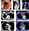
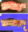
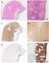
Case presentation: A 45-year-old woman with worsening dysphagia was transferred to our hospital. The patient was diagnosed with CES at 5 years of age and underwent surgery at another hospital. The patient underwent esophageal dilatation for stenosis at 36 years of age. Esophagoscopy performed at our hospital revealed a circumferential ulcerated lesion and stenosis 15-29 cm from the incisors. Histological examination of the biopsy specimen revealed squamous cell carcinoma. Computed tomography (CT) revealed abnormal circumferential wall thickening in parts of the cervical and almost the entire thoracic esophagus. 18F-fluorodeoxyglucose-positron emission tomography-CT revealed increased uptake in the cervical and upper esophagus. No uptake was observed in the muscular layers of the middle or lower esophagus. Based on these findings, the patient was diagnosed with clinical stage IVB cervical and upper esophageal cancer (T3N1M1 [supraclavicular lymph nodes]). The patient underwent a total esophagectomy after neoadjuvant chemotherapy. The esophagus was markedly thickened and tightly adhered to the adjacent organs. Severe fibrosis was observed around the trachea. Marked thickening of the muscular layer was observed throughout the esophagus; histopathological examination revealed that this thickening was due to increased smooth muscle mass. No cartilage, bronchial epithelium, or glands were observed. The carcinoma extended from the cervical to the middle esophagus, oral to the stenotic region. Finally, we diagnosed the patient with esophageal cancer developing on CES of the fibromuscular thickening type.
Conclusions: Chronic mechanical and chemical irritations are believed to cause cancer of the upper esophagus oral to a persistent CES, suggesting the need for long-term surveillance that focuses on residual stenosis and cancer development in patients with CES.
Keywords: Adult; Congenital esophageal stenosis; Esophageal cancer; Fibromuscular thickening.
© 2024. The Author(s).
Conflict of interest statement
The authors declare that they have no conflicts of interest.
Figures


Similar articles
-
Adult case of squamous cell carcinoma arising on congenital esophageal stenosis due to fibromuscular hypertrophy.Dis Esophagus. 2002;15(4):336-9. doi: 10.1046/j.1442-2050.2002.00270.x. Dis Esophagus. 2002. PMID: 12472484
-
Hybrid surgical approach for a large schwannoma from the cervical esophagus to the upper thoracic esophagus: a case report.Gen Thorac Cardiovasc Surg Cases. 2024 Oct 16;3(1):45. doi: 10.1186/s44215-024-00171-5. Gen Thorac Cardiovasc Surg Cases. 2024. PMID: 39516926 Free PMC article.
-
Endoscopic management of congenital esophageal stenosis.J Pediatr Surg. 2011 May;46(5):838-41. doi: 10.1016/j.jpedsurg.2011.02.010. J Pediatr Surg. 2011. PMID: 21616237
-
Management of congenital esophageal stenosis.J Pediatr Surg. 2002 Jul;37(7):1024-6. doi: 10.1053/jpsu.2002.33834. J Pediatr Surg. 2002. PMID: 12077763 Review.
-
[Congenital esophageal stenosis owing to ectopic tracheobronchial remnants: report of four cases and review of the literature].Zhonghua Er Ke Za Zhi. 2012 Aug;50(8):571-4. Zhonghua Er Ke Za Zhi. 2012. PMID: 23158732 Review. Chinese.
References
-
- Mochizuki K, Yokoi A, Urushihara N, Yabe K, Nakashima H, Kitagawa N, et al. Characteristics and treatment of congenital esophageal stenosis: a retrospective collaborative study from three Japanese children’s hospitals. J Pediatr Surg. 2021;56:1771–1775. doi: 10.1016/j.jpedsurg.2020.12.029. - DOI - PubMed
LinkOut - more resources
Full Text Sources

