

Multi-b-value DWI to evaluate the synergistic antiproliferation and anti-heterogeneity effects of bufalin plus sorafenib in an orthotopic HCC model
- PMID: 38467904
- PMCID: PMC10928042
- DOI: 10.1186/s41747-024-00448-y
Multi-b-value DWI to evaluate the synergistic antiproliferation and anti-heterogeneity effects of bufalin plus sorafenib in an orthotopic HCC model
Abstract
Background: Multi-b-value diffusion-weighted imaging (DWI) with different postprocessing models allows for evaluating hepatocellular carcinoma (HCC) proliferation, spatial heterogeneity, and feasibility of treatment strategies. We assessed synergistic effects of bufalin+sorafenib in orthotopic HCC-LM3 xenograft nude mice by using intravoxel incoherent motion (IVIM), diffusion kurtosis imaging (DKI), a stretched exponential model (SEM), and a fractional-order calculus (FROC) model.
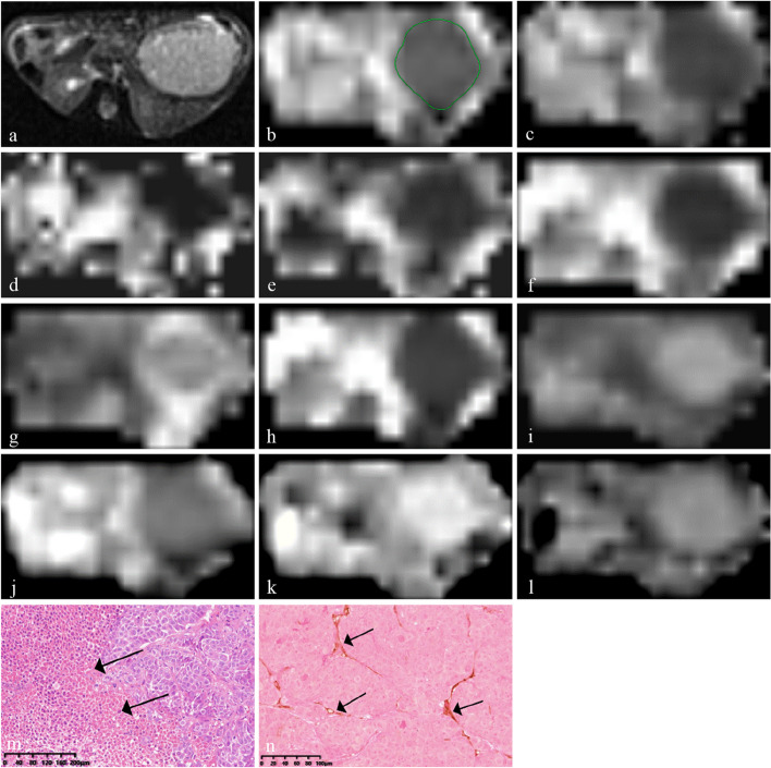
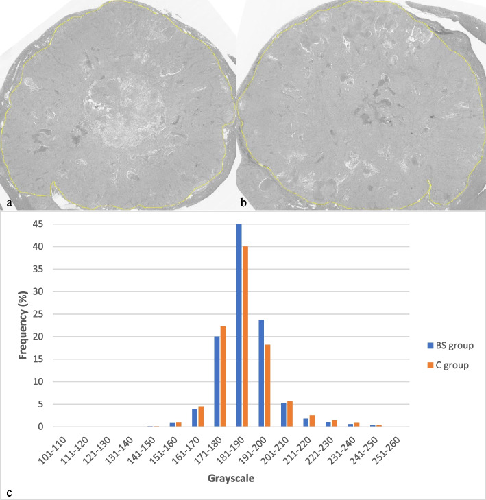
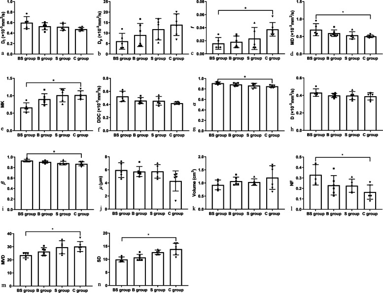
Methods: Twenty-four orthotopic HCC-LM3 xenograft mice were divided into bufalin+sorafenib, bufalin, sorafenib treatment groups, and a control group. Multi-b-value DWI was performed using a 3-T scanner after 3 weeks' treatment to obtain true diffusion coefficient Dt, pseudo-diffusion coefficient Dp, perfusion fraction f, mean diffusivity (MD), mean kurtosis (MK), distributed diffusion coefficient (DDC), heterogeneity index α, diffusion coefficient D, fractional order parameter β, and microstructural quantity μ. Necrotic fraction (NF), standard deviation (SD) of hematoxylin-eosin staining, and microvessel density (MVD) of anti-CD31 staining were evaluated. Correlations of DWI parameters with histopathological results were analyzed, and measurements were compared among four groups.
Results: In the final 22 mice, f positively correlated with MVD (r = 0.679, p = 0.001). Significantly good correlations of MK (r = 0.677), α (r = -0.696), and β (r= -0.639) with SD were observed (all p < 0.010). f, MK, MVD, and SD were much lower, while MD, α, β, and NF were higher in bufalin plus sorafenib group than control group (all p < 0.050).
Conclusion: Evaluated by IVIM, DKI, SEM, and FROC, bufalin+sorafenib was found to inhibit tumor proliferation and angiogenesis and reduce spatial heterogeneity in HCC-LM3 models.
Relevance statement: Multi-b-value DWI provides potential metrics for evaluating the efficacy of treatment in HCC.
Key points: • Bufalin plus sorafenib combination may increase the effectiveness of HCC therapy. • Multi-b-value DWI depicted HCC proliferation, angiogenesis, and spatial heterogeneity. • Multi-b-value DWI may be a noninvasive method to assess HCC therapeutic efficacy.
Keywords: Bufalin; Carcinoma (hepatocellular); Diffusion magnetic resonance imaging; Liver neoplasms; Sorafenib.
© 2024. The Author(s).
Conflict of interest statement
CF and ML are employed by Siemens Healthineer and provided MR technical support for this study under a Siemens collaboration agreement. They did not receive payment for participating in the study and had no personal motivation for the study outcome. The remaining authors declared no potential conflicts of interest concerning this article’s research, authorship, and/or publication.
Figures
References
MeSH terms
Substances
Grants and funding
- WL-XJRY-2021006K/Traditional Chinese Medicine Science and Technology Development Project of Shanghai Medical Innovation & Development Foundation
- WL-HBQN-2022009K/Traditional Chinese Medicine Science and Technology Development Project of Shanghai Medical Innovation & Development Foundation
- SHDC22022310-A/Shanghai Municipal Hospital Radiology Specialist Alliance
- SHDC22022310-B/Shanghai Municipal Hospital Radiology Specialist Alliance
LinkOut - more resources
Full Text Sources
Medical
Research Materials