Review
. 2024 Mar 11;25(1):121.
doi: 10.1186/s12931-024-02746-w.

Cardiopulmonary exercise test to detect cardiac dysfunction from pulmonary vascular disease

Affiliations
Review

Cardiopulmonary exercise test to detect cardiac dysfunction from pulmonary vascular disease

Mona Alotaibi et al. Respir Res. .

Abstract

Background: Cardiac dysfunction from pulmonary vascular disease causes characteristic findings on cardiopulmonary exercise testing (CPET). We tested the accuracy of CPET for detecting inadequate stroke volume (SV) augmentation during exercise, a pivotal manifestation of cardiac limitation in patients with pulmonary vascular disease.

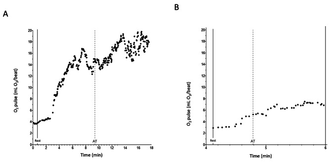
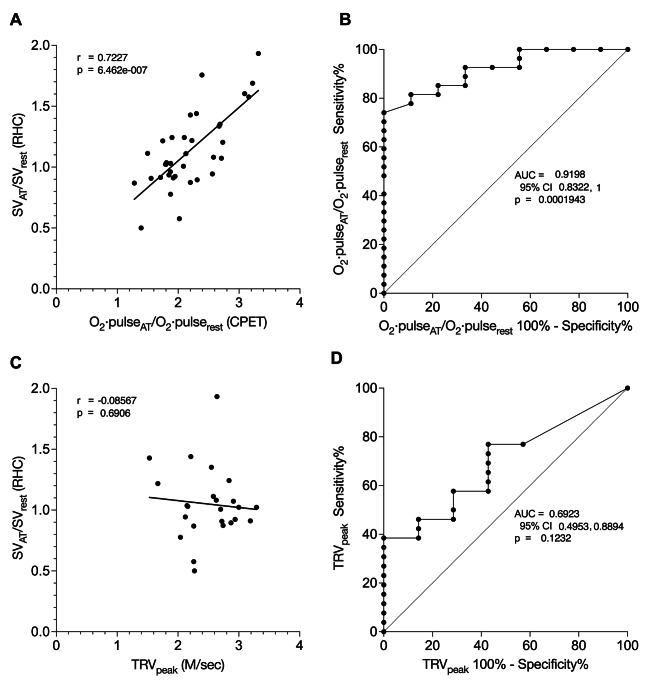
Methods: We reviewed patients with suspected pulmonary vascular disease in whom CPET and right heart catheterization (RHC) measurements were taken at rest and at anaerobic threshold (AT). We correlated CPET-determined O2·pulseAT/O2·pulserest with RHC-determined SVAT/SVrest. We evaluated the sensitivity and specificity of O2·pulseAT/O2·pulserest to detect SVAT/SVrest below the lower limit of normal (LLN). For comparison, we performed similar analyses comparing echocardiographically-measured peak tricuspid regurgitant velocity (TRVpeak) with SVAT/SVrest.

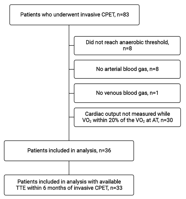
Results: From July 2018 through February 2023, 83 simultaneous RHC and CPET were performed. Thirty-six studies measured O2·pulse and SV at rest and at AT. O2·pulseAT/O2·pulserest correlated highly with SVAT/SVrest (r = 0.72, 95% CI 0.52, 0.85; p < 0.0001), whereas TRVpeak did not (r = -0.09, 95% CI -0.47, 0.33; p = 0.69). The AUROC to detect SVAT/SVrest below the LLN was significantly higher for O2·pulseAT/O2·pulserest (0.92, SE 0.04; p = 0.0002) than for TRVpeak (0.69, SE 0.10; p = 0.12). O2·pulseAT/O2·pulserest of less than 2.6 was 92.6% sensitive (95% CI 76.6%, 98.7%) and 66.7% specific (95% CI 35.2%, 87.9%) for deficient SVAT/SVrest.

Conclusions: CPET detected deficient SV augmentation more accurately than echocardiography. CPET-determined O2·pulseAT/O2·pulserest may have a prominent role for noninvasive screening of patients at risk for pulmonary vascular disease, such as patients with persistent dyspnea after pulmonary embolism.

Keywords: Cardiopulmonary exercise test (CPET); Echocardiography; Pulmonary embolism; Pulmonary vascular disease; Stroke volume augmentation.


Conflict of interest statement

Summary of conflict-of-interest statements: The authors report no conflicts of interest and no financial support relevant to this work.

Figures

Fig. 1
Fig. 1
O2·pulse at rest (O2·pulserest) and at anaerobic threshold (O2·pulseAT). (A) Normal increase in O2·pulse from rest (O2·pulserest, solid vertical line) to anaerobic threshold (O2·pulseAT, dashed vertical line). O2·pulserest was 4.5 ml O2/beat and O2·pulseAT was 14.5 ml O2/beat. (B) Pathologically low increase from O2·pulserest to O2·pulseAT. O2·pulserest was 3.8 ml O2/beat and O2·pulseAT was 7.3 ml O2/beat
Fig. 2
Fig. 2
Patient selection. CPET, cardiopulmonary exercise test; VO2, oxygen consumption; AT, anaerobic threshold; TTE, transthoracic echocardiography
Fig. 3
Fig. 3
O2·pulseAT/O2·pulserest compared to TRVpeak to predict stroke volume augmentation. A. O2·pulseAT/O2·pulserest determined from CPET data (see text) correlated with stroke volume augmentation from rest to anaerobic threshold (SVAAT) measured by right heart catheterization (p < 0.0001). B. The ROC curve of O2·pulseAT/O2·pulserest to detect SVAAT below the lower limit of normal (LLN) had an area under the ROC curve of 0.92 (SE 0.04, p = 0.0002). C. Tricuspid regurgitant velocity peak (TRVpeak) measured by echocardiography did not correlate with SVAAT (p = 0.69). D. The ROC curve of TRVpeak to detect SVAAT below the LLN had an area of 0.69 (SE 0.10), which was not statistically significant (p = 0.12)

Similar articles

Cited by

References

    1. Morris TA, Fernandes TM, Channick R. How we do it: evaluation of dyspnea and exercise intolerance after acute pulmonary embolism. Chest. - PMC - PubMed
    1. Farmakis IT, Valerio L, Barco S et al. Cardiopulmonary exercise testing in the follow-up after acute pulmonary embolism. Eur Respiratory Journal: Official J Eur Soc Clin Respiratory Physiol 2023. - PMC - PubMed
    1. Holverda S, Gan CT, Marcus JT, Postmus PE, Boonstra A, Vonk-Noordegraaf A. Impaired stroke volume response to exercise in pulmonary arterial hypertension. J Am Coll Cardiol. 2006;47(8):1732–3. doi: 10.1016/j.jacc.2006.01.048. - DOI - PubMed
    1. Surie S, van der Plas MN, Marcus JT, et al. Effect of pulmonary endarterectomy for chronic thromboembolic pulmonary hypertension on stroke volume response to exercise. Am J Cardiol. 2014;114(1):136–40. doi: 10.1016/j.amjcard.2014.04.016. - DOI - PubMed
    1. Fernandes TM, Alotaibi M, Strozza DM, et al. Dyspnea Postpulmonary Embolism from physiological Dead Space Proportion and Stroke volume defects during Exercise. Chest. 2020;157(4):936–44. doi: 10.1016/j.chest.2019.10.047. - DOI - PubMed