. 2024 Mar 11;19(1):46.
doi: 10.1186/s13020-024-00914-0.

Salvianolic acid C attenuates cerebral ischemic injury through inhibiting neuroinflammation via the TLR4-TREM1-NF-κB pathway

Affiliations

Salvianolic acid C attenuates cerebral ischemic injury through inhibiting neuroinflammation via the TLR4-TREM1-NF-κB pathway

Wenbo Guo et al. Chin Med. .

Abstract

Background: Stroke is a leading cause of mortality and disability with ischemic stroke being the most common type of stroke. Salvianolic acid C (SalC), a polyphenolic compound found in Salviae Miltiorrhizae Radix et Rhizoma, has demonstrated therapeutic potential in the recovery phase of ischemic stroke. However, its pharmacological effects and underlying mechanisms during the early stages of ischemic stroke remain unclear. This study aimed to examine the potential mechanism of action of SalC during the early phase of ischemic stroke using network pharmacology strategies and RNA sequencing analysis.

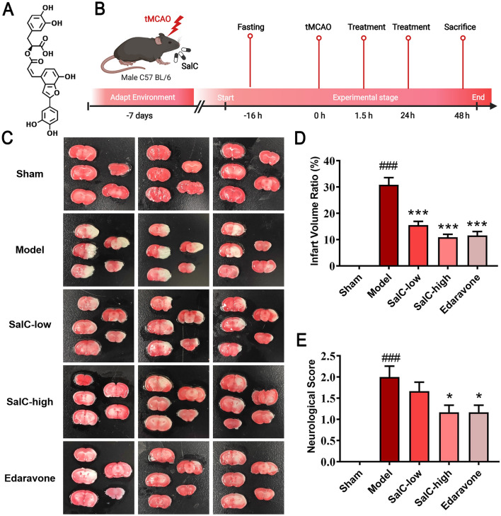
Methods: SalC effects on infarct volume, neurological deficits, and histopathological changes were assessed in a mouse model of transient middle cerebral artery occlusion (tMCAO). By integrating RNA sequencing data with a cerebral vascular disease (CVD)-related gene database, a cerebral ischemic disease (CID) network containing dysregulated genes from the tMCAO model was constructed. Network analysis algorithms were applied to evaluate the key nodes within the CID network. In vivo and in vitro validation of crucial targets within the identified pathways was conducted.

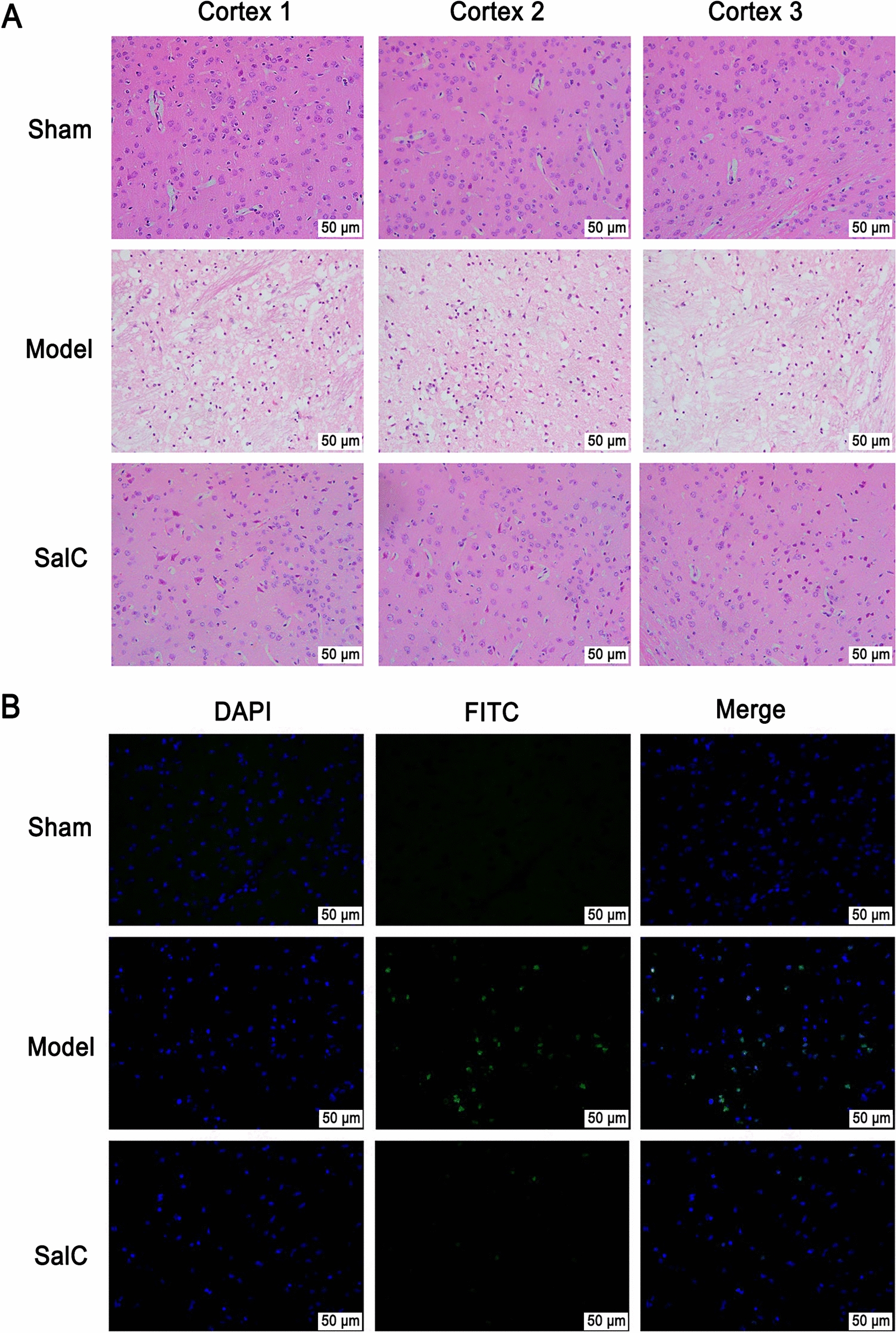
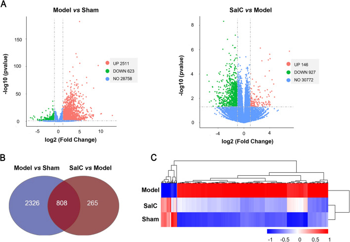
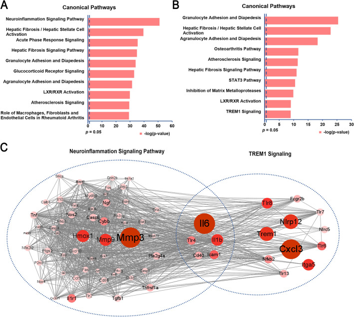
Results: SalC treatment significantly reduced infarct volume, improved neurological deficits, and reversed pathological changes in the tMCAO mouse model. The integration of RNA sequencing data revealed an 80% gene reversion rate induced by SalC within the CID network. Among the reverted genes, 53.1% exhibited reversion rates exceeding 50%, emphasizing the comprehensive rebalancing effect of SalC within the CID network. Neuroinflammatory-related pathways regulated by SalC, including the toll-like-receptor 4 (TLR4)- triggering receptor expressed on myeloid cells 1 (TREM1)-nuclear factor kappa B (NF-κB) pathway, were identified. Further in vivo and in vitro experiments confirmed that TLR4-TREM1-NF-κB pathway was down-regulated by SalC in microglia, which was essential for its anti-inflammatory effect on ischemic stroke.

Conclusions: SalC attenuated cerebral ischemic injury by inhibiting neuroinflammation mediated by microglia, primarily through the TLR4-TREM1-NF-κB pathway. These findings provide valuable insights into the potential therapeutic benefits of SalC in ischemic stroke.

Keywords: Cerebral ischemic stroke; Disease network; Neuroinflammation; RNA transcriptome sequencing; Salvianolic acid C; TREM1.


Conflict of interest statement

The authors declare no competing interests.

Figures

Fig. 1
Fig. 1
SalC reduces cerebral infarction and improves neurological function of tMCAO mice. A Structural formula of SalC. B Experimental scheme. C Representative TTC-stained coronary brain sections (white, infarcted area; red, non-infarcted area). D Statistics of the volume ratio of cerebral infarction in mice. E Scores of neurological deficits in different groups. Data are expressed as mean ± SEM, n = 6. The significance was determined by one-way ANOVA followed by Dunnett's posterior analysis, ###P < 0.001, versus the sham group; *P < 0.05, ***P < 0.001, versus the model group
Fig. 2
Fig. 2
SalC reduces brain damage and neuron apoptosis in tMCAO mice. A H&E staining of three areas in the penumbra of the brain cortex. B TUNEL staining with representative photos showed apoptotic neurons in the penumbra of ischemic cortex (appearing yellow-green due to FITC labeling). n = 3, magnification of 400 ×
Fig. 3
Fig. 3
Transcriptional analysis shows that SalC restores gene expression in the brain tissues of tMCAO mice. A Volcano plot shows the DEGs. Genes with absolute fold changes > 2 and P value < 0.05 were highlighted in red and green, indicating up- and down-regulated genes, respectively. B Venn diagram of the DEGs. C Hierarchical clustering analysis of the 808 genes that were dysregulated under the modeling of tMCAO and reversed by SalC
Fig. 4
Fig. 4
SalC recovers the unbalanced CID network disturbed by tMCAO modeling. A CID network. Node size represents NTRA rank. B Unbalanced CID network disturbed by tMCAO modeling. Red indicates log2 Fold Change (model/sham) > 0, representing upregulation of this gene expression in the model group compared with the sham group; Green indicates log2 Fold Change (model/sham) < 0, representing downregulation of this gene expression in the model group compared with the sham group. Moreover, the shade of the color indicates the degree of upregulation and downregulation. C Effect of SalC on the regulation of CID network. Blue represents the efficiency of EoR > 0, the darker the color, the greater the degree of regulation; Green represents EoR < 0, indicating that SalC has no regulatory effect on this gene. n = 4
Fig. 5
Fig. 5
SalC mitigates stroke in mice by suppressing neuroinflammation and TREM1 signaling after tMCAO-induced cerebral ischemia. A The top 10 canonical pathways involved in the top 300 NTRA-ranked genes with EoR > 0. B The top 10 canonical pathways involved in the 808 DEGs. C Diagram of gene interactions in neuroinflammation and TREM1 signaling pathways. Node size and color shades represent the size of |log2 Fold Change|
Fig. 6
Fig. 6
SalC attenuates cerebral ischemic stroke in tMCAO mice by inhibiting neuroinflammation via the TLR4-TREM1-NF-κB pathway. A The gene expressions of Trem1, Il6, and Cxcl1 detected by qRT-PCR to validate transcriptome sequencing results. β-actin was used as an internal reference. B The protein expression levels of TREM1, TLR4, p-p65, and p65 in brain tissue were analyzed by western blot. β-actin was used as an internal standard. C Quantification was performed by Image Lab software. Data are expressed as mean ± SEM, and differences between groups were determined by t-test, n = 4. ###P < 0.001, #P < 0.05 versus the sham group; *P < 0.05, versus the model group
Fig. 7
Fig. 7
SalC inhibits TREM1 expression in microglia. Representative double-staining immunofluorescence of Iba-1 (red) and TREM1 (green) in the ischemic transition region of brain sections. Nuclei were stained with DAPI (blue). n = 3, magnification 200 ×
Fig. 8
Fig. 8
The protective effect of SalC on OGD/R injury in vitro. A Effects of SalC on OGD/R modeling of BV2 cells. B Effects of SalC on OGD/R modeling of HT22 cells. C Effects of SalC on the co-culture system of BV2 and HT22 cells. D The protein expression levels of TREM1, TLR4, p-p65 and p65 in BV2 cells were analyzed by western blot. β-actin was used as an internal standard. E Quantification was performed by Image Lab software. Data are expressed as mean ± SEM, and differences between groups were determined by t-test, n = 3. ###P < 0.001, ##P < 0.01, #P < 0.05, versus the sham group; ***P < 0.001, **P < 0.01, *P < 0.05, versus the model group
Fig. 9
Fig. 9
The scheme of the mechanism for the protective effects of SalC on cerebral ischemia stroke in tMCAO mice by inhibiting neuroinflammation via the TLR4-TREM1-NF-κB pathway

References

    1. Feigin VL, Norrving B, Mensah GA. Global burden of stroke. Circ Res. 2017;120(3):439–448. doi: 10.1161/CIRCRESAHA.116.308413. - DOI - PubMed
    1. Virani SS, Alonso A, Benjamin EJ, Bittencourt MS, Callaway CW, Carson AP, Chamberlain AM, Chang AR, Cheng S, Delling FN, et al. Heart Disease and Stroke Statistics-2020 Update: a report from the American Heart Association. Circulation. 2020;141(9):e139–e596. doi: 10.1161/CIR.0000000000000757. - DOI - PubMed
    1. Valery L, Benjamin A. Global, regional, and national burden of stroke and its risk factors, 1990–2019: a systematic analysis for the Global Burden of Disease Study 2019. Lancet Neurol. 2021;20(10):795–820. doi: 10.1016/S1474-4422(21)00252-0. - DOI - PMC - PubMed
    1. Feigin VL, Nguyen G, Cercy K, Johnson CO, Alam T, Parmar PG, Abajobir AA, Abate KH, Abd-Allah F, Abejie AN, et al. Global, regional, and country-specific lifetime risks of stroke, 1990 and 2016. New Engl J Med. 2018;379(25):2429–2437. doi: 10.1056/NEJMoa1804492. - DOI - PMC - PubMed
    1. Hlavica M, Diepers M, Garcia-Esperon C, Ineichen BV, Nedeltchev K, Kahles T, Remonda L. Pharmacological recanalization therapy in acute ischemic stroke—evolution, current state and perspectives of intravenous and intra-arterial thrombolysis. J Neuroradiol. 2015;42(1):30–46. doi: 10.1016/j.neurad.2014.11.004. - DOI - PubMed

LinkOut - more resources