

3D-printed bredigite scaffolds with ordered arrangement structures promote bone regeneration by inducing macrophage polarization in onlay grafts
- PMID: 38468312
- PMCID: PMC10926610
- DOI: 10.1186/s12951-024-02362-2
3D-printed bredigite scaffolds with ordered arrangement structures promote bone regeneration by inducing macrophage polarization in onlay grafts
Abstract
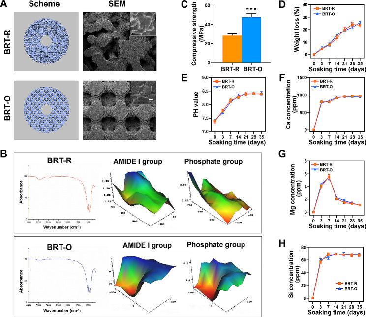
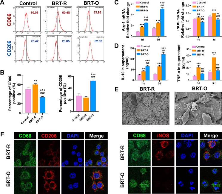
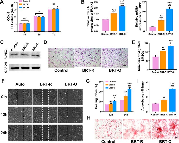
Bone tissue engineering scaffolds may provide a potential strategy for onlay bone grafts for oral implants. For determining the fate of scaffold biomaterials and osteogenesis effects, the host immune response is crucial. In the present study, bredigite (BRT) bioceramic scaffolds with an ordered arrangement structure (BRT-O) and a random morphology (BRT-R) were fabricated. The physicochemical properties of scaffolds were first characterized by scanning electron microscopy, mechanical test and micro-Fourier transform infrared spectroscopy. In addition, their osteogenic and immunomodulatory properties in an onlay grafting model were investigated. In vitro, the BRT-O scaffolds facilitated the macrophage polarization towards a pro-regenerative M2 phenotype, which subsequently facilitated the migration and osteogenic differentiation of bone marrow-derived mesenchymal stem cells. In vivo, an onlay grafting model was successfully established in the cranium of rabbits. In addition, the BRT-O scaffolds grafted on rabbit cranium promoted bone regeneration and CD68 + CD206 + M2 macrophage polarization. In conclusion, the 3D-printed BRT-O scaffold presents as a promising scaffold biomaterial for onlay grafts by regulating the local immune microenvironment.
Keywords: Bone regeneration; Bone scaffold; Onlay graft; Oral implant; Osteoimmunomodulation.
© 2024. The Author(s).
Conflict of interest statement
The authors declare no competing interests.
Figures

Similar articles
-
The 3D-Printed Ordered Bredigite Scaffold Promotes Pro-Healing of Critical-Sized Bone Defects by Regulating Macrophage Polarization.Int J Nanomedicine. 2023 Feb 20;18:917-932. doi: 10.2147/IJN.S393080. eCollection 2023. Int J Nanomedicine. 2023. PMID: 36844434 Free PMC article.
-
Three-Dimensional-Printed Ordered Bredigite Scaffolds with Dual Bioactivities Promote Osteochondral Regeneration.ACS Appl Mater Interfaces. 2025 Jul 23;17(29):41707-41718. doi: 10.1021/acsami.5c07388. Epub 2025 Jul 14. ACS Appl Mater Interfaces. 2025. PMID: 40660801
-
Osteoimmunomodulation unveiled: Enhancing bone regeneration with 3D-printed PLLA/β-TCP/CS scaffolds.Colloids Surf B Biointerfaces. 2025 Aug;252:114674. doi: 10.1016/j.colsurfb.2025.114674. Epub 2025 Apr 1. Colloids Surf B Biointerfaces. 2025. PMID: 40186926
-
3D-printed bioceramic scaffolds: From bone tissue engineering to tumor therapy.Acta Biomater. 2018 Oct 1;79:37-59. doi: 10.1016/j.actbio.2018.08.026. Epub 2018 Aug 28. Acta Biomater. 2018. PMID: 30165201 Review.
-
Chitosan-based biomaterials promote bone regeneration by regulating macrophage fate.J Mater Chem B. 2024 Aug 7;12(31):7480-7496. doi: 10.1039/d3tb02563b. J Mater Chem B. 2024. PMID: 39016095 Review.
Cited by
-
Magnesium malate-modified calcium phosphate bone cement promotes the repair of vertebral bone defects in minipigs via regulating CGRP.J Nanobiotechnology. 2024 Jun 25;22(1):368. doi: 10.1186/s12951-024-02595-1. J Nanobiotechnology. 2024. PMID: 38918787 Free PMC article.
-
Advances in the application and research of biomaterials in promoting bone repair and regeneration through immune modulation.Mater Today Bio. 2024 Dec 16;30:101410. doi: 10.1016/j.mtbio.2024.101410. eCollection 2025 Feb. Mater Today Bio. 2024. PMID: 39811613 Free PMC article. Review.
-
3D-printed bioceramic scaffolds for bone defect repair: bone aging and immune regulation.Front Bioeng Biotechnol. 2025 Mar 31;13:1557203. doi: 10.3389/fbioe.2025.1557203. eCollection 2025. Front Bioeng Biotechnol. 2025. PMID: 40242352 Free PMC article. Review.
References
-
- Severi M, Simonelli A, Farina R, Tu YK, Lan CH, Shih MC, Trombelli L. Effect of lateral bone augmentation procedures in correcting peri-implant bone dehiscence and fenestration defects: a systematic review and network meta-analysis. Clin Implant Dent Relat Res. 2022;24:251–64. doi: 10.1111/cid.13078. - DOI - PMC - PubMed
MeSH terms
Substances
Grants and funding
LinkOut - more resources
Full Text Sources
