. 2024 Mar 12;15(1):43.
doi: 10.1186/s40104-024-01002-x.

In-depth proteome characterization of endometrium and extraembryonic membranes during implantation in pig

Affiliations

In-depth proteome characterization of endometrium and extraembryonic membranes during implantation in pig

Maria A Gil et al. J Anim Sci Biotechnol. .

Abstract

Background: Proteome characterization of the porcine endometrium and extraembryonic membranes is important to understand mother-embryo cross-communication. In this study, the proteome of the endometrium and chorioallantoic membrane was characterized in pregnant sows (PS) during early gestation (d 18 and 24 of gestation) and in the endometrium of non-pregnant sows (NPS) during the same days using LC-MS/MS analysis. The UniProtKB database and ClueGO were used to obtain functional Gene Ontology annotations and biological and functional networks, respectively.

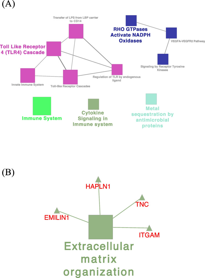
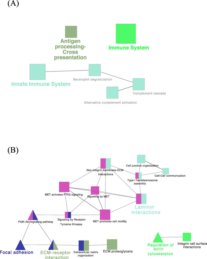
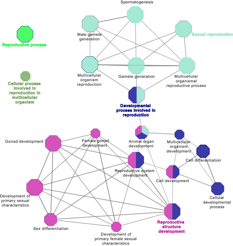
Results: Our analysis yielded 3,254 and 3,457 proteins identified in the endometrium of PS and NPS, respectively; of these, 1,753 being common while 1,501 and 1,704 were exclusive to PS and NPS, respectively. In addition, we identified 3,968 proteins in the extraembryonic membranes of PS. Further analyses of function revealed some proteins had relevance for the immune system process and biological adhesion in endometrium while the embryonic chorion displayed abundance of proteins related to cell adhesion and cytoskeletal organization, suggesting they dominated the moment of endometrial remodeling, implantation and adhesion of the lining epithelia. Data are available via ProteomeXchange with identifier PXD042565.

Conclusion: This is the first in-depth proteomic characterization of the endometrium and extraembryonic membranes during weeks 3 to 4 of gestation; data that contribute to the molecular understanding of the dynamic environment during this critical period, associated with the majority of pregnancy losses.

Keywords: Endometrium; Extraembryonic membranes; Implantation; Pig; Proteome.


Conflict of interest statement

The authors declare that they have no competing interests.

Figures

Fig. 1
Fig. 1
Gene Ontology analysis of the endometrial proteome of pregnant sows during implantation. A Biological process. B Reproductive process. C Molecular function. D Cellular component
Fig. 2
Fig. 2
Gene Ontology analysis of the endometrial proteome of non-pregnant sows at d 18 and 24 of the cycle. A Biological process. B Reproductive process. C Molecular function. D Cellular component
Fig. 3
Fig. 3
Visualization of the reactome pathways analysis of unique proteins involved in immune system process (A) and biological adhesion annotation (B) in pregnant sows. The size of the nodes indicates the pathway´s enrichment significance. The following ClueGO parameters were used: P-value cut-off = 0.05; Statistical test used the enrichment/depletion (two-sided hypergeometric test); Bonferroni step down with minimum GO level 2; maximum GO level 6; kappa score threshold equal to 0.4. GO = Gene Ontology. Network (A) includes the interaction of CCR5, HMOX1, IFI35, ISG15, LBP, MAP2K1, MAPK14, SLA, STXBP2, VAMP7, CD14, DNM1, LBP, MAP2K1, MAPK14 and S100A9 proteins
Fig. 4
Fig. 4
Visualization of the biological gene ontology analysis of unique proteins involved in the reproductive process annotation in pregnant sows. The size of the nodes indicates the pathway´s enrichment significance. The following ClueGO parameters were used: P-value cut-off = 0.001; Statistical test used the enrichment/depletion (two-sided hypergeometric test); Bonferroni step down with minimum GO level 2; maximum GO level 6; kappa score threshold equal to 0.4. GO = Gene Ontology. Network include the involvement of HECTD1, HSD17B2, MAP2K1, MAPK14, RPS6, MSH2, DLG1 and INHBB proteins among others
Fig. 5
Fig. 5
Visualization of the reactome pathways analysis of unique proteins involved in immune system process (A) and in biological adhesion annotation (B) in non-pregnant sows. The size of the nodes indicates the pathway´s enrichment significance. The following ClueGO parameters were used: P-value cut-off = 0.05; Statistical test used the enrichment/depletion (two-sided hypergeometric test); Bonferroni step down with minimum GO level 2; maximum GO level 6; kappa score threshold equal to 0.4. GO = Gene Ontology. Network (A) includes the interaction of ABI1, C5AR1, CD44, CFD, CFP, ELANE, HP, HRAS, IFIH1, MMP9, MYD88, PSMB6 and VNN1 proteins. Network (B) includes the interaction of FN1, ITGA3, ITGA9, ITGB4, LAMA3, LAMA5, THBS4, TNXB, EGFR, ITGAX, EGFR, PARVA, and TNXB proteins
Fig. 6
Fig. 6
Visualization of the biological gene ontology analysis of unique proteins involved in the reproductive process annotation in non-pregnant sows. The size of the nodes indicates the pathway’s enrichment significance. The following ClueGO parameters were used: P-value cut-off = 0.001; Statistical test used the enrichment/depletion (two-sided hypergeometric test); Bonferroni step down with minimum GO level 2; maximum GO level 6; kappa score threshold equal to 0.4. GO = Gene Ontology
Fig. 7
Fig. 7
Gene Ontology analysis of the extraembryonic membranes proteome of pregnant sows at d 18 and 24 of gestation. A Biological process. B Reproductive process. C Molecular function. D Cellular component
Fig. 8
Fig. 8
Visualization of the functional analysis of the proteins involved in the reproductive process annotation in extraembryonic membranes of pregnant sows at d 18 and 24 of gestation. The size of the nodes indicates the pathway´s enrichment significance. The following ClueGO parameters were used: P-value cut-off = 0.001; Statistical test used the enrichment/depletion (two-sided hypergeometric test); Bonferroni step down with minimum GO level 2; maximum GO level 6; kappa score threshold equal to 0.4. GO = Gene Ontology. Network includes the interaction of RAD21, SMC1A, SMC3, CCT3, CCT4, CCT8 and TCP1 proteins
Fig. 9
Fig. 9
Visualization of the biological gene ontology analysis of the proteins involved in the reproductive process annotation in extraembryonic membranes of pregnant sows at d 18 and 24 of gestation. The size of the nodes indicates the pathway´s enrichment significance. The following ClueGO parameters were used: P-value cut-off = 0.001; Statistical test used the enrichment/depletion (two-sided hypergeometric test); Bonferroni step down with minimum GO level 2; maximum GO level 6; kappa score threshold equal to 0.4. GO = Gene Ontology. Network includes the interaction of CCDC134, HSD17B2, HSP90AB1, KRT19, MAPK14, NOTCH2, PRDX3, PTGS2, RTCB, ST14, ACE, APOB, BAX, CCDC134, CDK1, CELF1, CTNNB1, DHX36, DLG1, HSD17B2, ITGB4, KDR, KRT19, LRP2, MSH2, NOTCH1, NOTCH2, PAFAH1B1, PDGFRA, PRKACA, RAD23B, RTCB, SEPTIN7, SMAD1 and ST14 proteins
Fig. 10
Fig. 10
Representative validation by Western blot of HMOX1 (hemo oxidase 1) and RPS6 (40S ribosomal protein) proteins in the porcine endometrium of pregnant sows (a) of PTN protein (pleiotrophin) in the porcine endometrium of non-pregnant sows (b) and THBS1 protein (thrombospondin1) in the extraembryonic membranes of pregnant sows (c)

References

    1. Almiñana C, Heath P, Wilkinson S, Sanchez-Osorio J, Cuello C, Parrilla I, et al. Early developing pig embryos mediate their own environment in the maternal tract. PLoS One. 2012;7(3):33625. doi: 10.1371/journal.pone.0033625. - DOI - PMC - PubMed
    1. Fleming TP, Kwong WY, Porter R, Ursell E, Fesenko I, Wilkins A, et al. The embryo and its future. Biol Reprod. 2004;71(4):1046–1054. doi: 10.1095/biolreprod.104.030957. - DOI - PubMed
    1. Pope WF, First NL. Factors affecting the survival of pig embryos. Theriogenology. 1985;23(1):91–105. doi: 10.1016/0093-691X(85)90075-5. - DOI
    1. Meyer AE, Pfeiffer CA, Brooks KE, Spate LD, Benne JA, Cecil R, Samuel MS, et al. New perspective on conceptus estrogens in maternal recognition and pregnancy establishment in the pig. Biol Reprod. 2019;101(1):148–161. doi: 10.1093/biolre/ioz058. - DOI - PubMed
    1. Edwards AK, Wessels JM, Kerr A, Tayade C. An overview of molecular and cellular mechanisms associated with porcine pregnancy success or failure. Reprod Domest Anim. 2012;47(SUPPL.4):394–401. doi: 10.1111/j.1439-0531.2012.02103.x. - DOI - PubMed