

RB1 loss induces quiescent state through downregulation of RAS signaling in mammary epithelial cells
- PMID: 38468443
- PMCID: PMC11093197
- DOI: 10.1111/cas.16122
RB1 loss induces quiescent state through downregulation of RAS signaling in mammary epithelial cells
Abstract
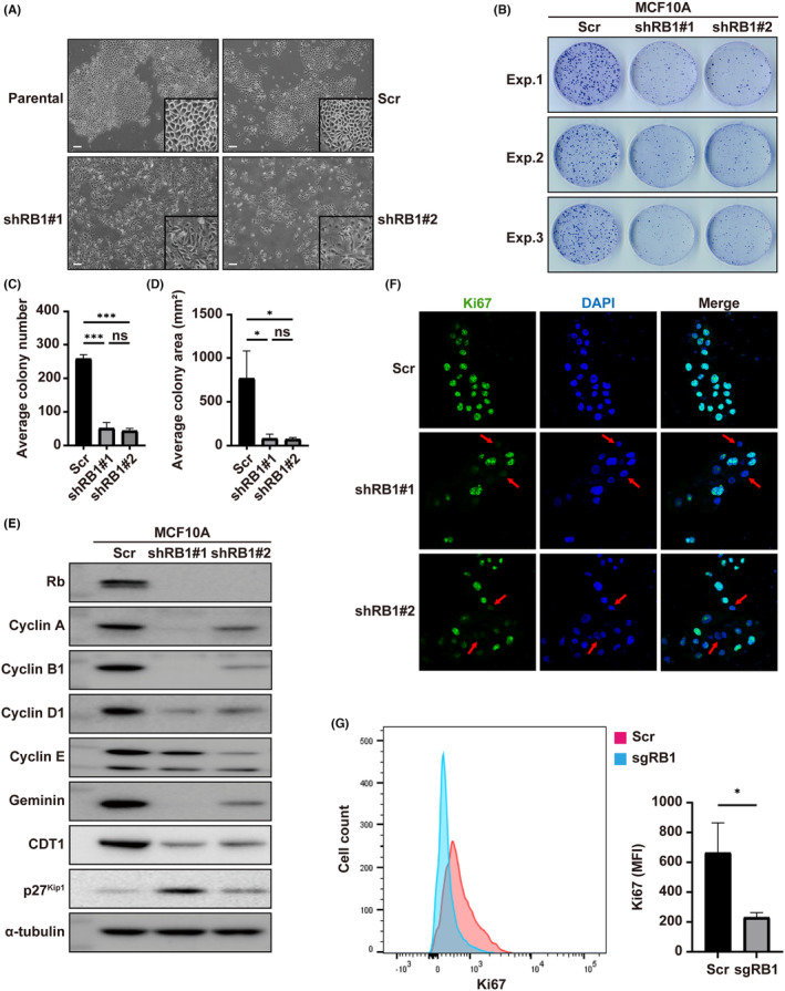
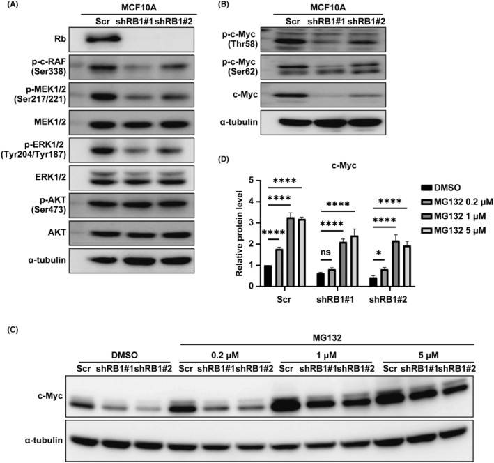
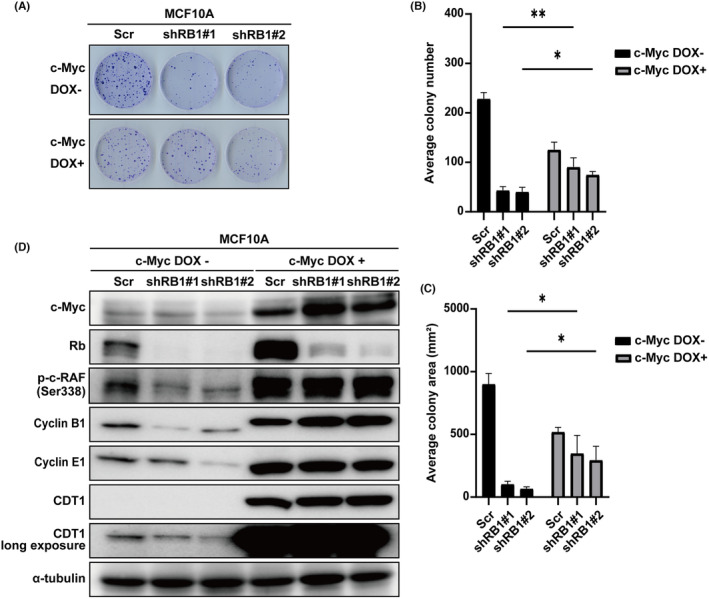
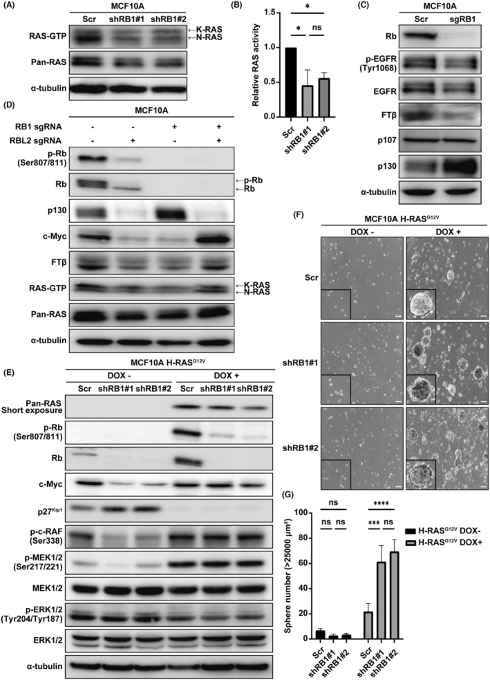
While loss of function (LOF) of retinoblastoma 1 (RB1) tumor suppressor is known to drive initiation of small-cell lung cancer and retinoblastoma, RB1 mutation is rarely observed in breast cancers at their initiation. In this study, we investigated the impact on untransformed mammary epithelial cells given by RB1 LOF. Depletion of RB1 in anon-tumorigenic MCF10A cells induced reversible growth arrest (quiescence) featured by downregulation of multiple cyclins and MYC, upregulation of p27KIP1, and lack of expression of markers which indicate cellular senescence or epithelial-mesenchymal transition (EMT). We observed a similar phenomenon in human mammary epithelial cells (HMEC) as well. Additionally, we found that RB1 depletion attenuated the activity of RAS and the downstream MAPK pathway in an RBL2/p130-dependent manner. The expression of farnesyltransferase β, which is essential for RAS maturation, was found to be downregulated following RB1 depletion also in an RBL2/p130-dependent manner. These findings unveiled an unexpected mechanism whereby normal mammary epithelial cells resist to tumor initiation upon RB1 LOF.
Keywords: MYC; RAS; RB1; RBL2/p130; quiescence.
© 2024 The Authors. Cancer Science published by John Wiley & Sons Australia, Ltd on behalf of Japanese Cancer Association.
Conflict of interest statement
The authors have no financial interest to disclose. Dr. Chiaki Takahashi is an associate editor of
Figures
Similar articles
-
RABL6A Is an Essential Driver of MPNSTs that Negatively Regulates the RB1 Pathway and Sensitizes Tumor Cells to CDK4/6 Inhibitors.Clin Cancer Res. 2020 Jun 15;26(12):2997-3011. doi: 10.1158/1078-0432.CCR-19-2706. Epub 2020 Feb 21. Clin Cancer Res. 2020. PMID: 32086342 Free PMC article.
-
Suppression of RAD21 Induces Senescence of MDA-MB-231 Human Breast Cancer Cells Through RB1 Pathway Activation Via c-Myc Downregulation.J Cell Biochem. 2016 Jun;117(6):1359-69. doi: 10.1002/jcb.25426. Epub 2015 Nov 20. J Cell Biochem. 2016. PMID: 26529363
-
EMT inducers catalyze malignant transformation of mammary epithelial cells and drive tumorigenesis towards claudin-low tumors in transgenic mice.PLoS Genet. 2012;8(5):e1002723. doi: 10.1371/journal.pgen.1002723. Epub 2012 May 24. PLoS Genet. 2012. PMID: 22654675 Free PMC article.
-
The significance of RB1 in multiple myeloma.Front Immunol. 2024 Nov 27;15:1415972. doi: 10.3389/fimmu.2024.1415972. eCollection 2024. Front Immunol. 2024. PMID: 39664374 Free PMC article. Review.
-
Targeting the untargetable: RB1-deficient tumours are vulnerable to Skp2 ubiquitin ligase inhibition.Br J Cancer. 2022 Oct;127(6):969-975. doi: 10.1038/s41416-022-01898-0. Epub 2022 Jun 25. Br J Cancer. 2022. PMID: 35752713 Free PMC article. Review.
References
-
- Shamma A, Takegami Y, Miki T, et al. Rb regulates DNA damage response and cellular senescence through E2F‐dependent suppression of N‐ras isoprenylation. Cancer Cell. 2009;15:255‐269. - PubMed
MeSH terms
Grants and funding
LinkOut - more resources
Full Text Sources
Molecular Biology Databases
Research Materials
Miscellaneous
