

A rare case of De Garengeot hernia: CT findings
- PMID: 38468719
- PMCID: PMC10927328
- DOI: 10.1093/bjrcr/uaae009
A rare case of De Garengeot hernia: CT findings
Abstract
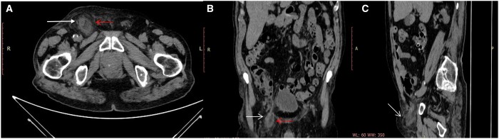
We report a case of "De Garengeot's hernia" (DGH), a rare condition that occurs when the inflamed appendix is localized inside a femoral hernia. The appendix may be involved in inflammatory or necrotic processes and the treatment is emergency surgery. It is usually discovered by chance during surgery. It occurs in 0.5%-5% of all femoral hernias. In 0.08%-0.13% of cases, the appendix can present inflammatory or necrotic processes due to the narrowness of the neck of the femoral canal; in these cases, an emergency surgery is required through a no standard surgical procedure. In the other cases, it is usually found accidentally during surgical repair of the hernia or more rarely diagnosed preoperatively by CT. Therefore, the purpose of our study is to report a case of DGH describing CT main findings in order to improve the preoperative diagnosis.
Keywords: Amyand hernia; De Garengeot hernia; appendicitis; appendix; computed tomography; femoral; hernia; inguinal.
© The Author(s) 2024. Published by Oxford University Press on behalf of the British Institute of Radiology.
Conflict of interest statement
The authors declare no conflict of interest.
Figures

References
Publication types
LinkOut - more resources
Full Text Sources
