[Preprint]. 2024 Feb 23:2024.02.20.581224.
doi: 10.1101/2024.02.20.581224.

p300 KAT regulates SOX10 stability and function in human melanoma

Affiliations

p300 KAT regulates SOX10 stability and function in human melanoma

Aaron Waddell et al. bioRxiv. .

Update in

Abstract

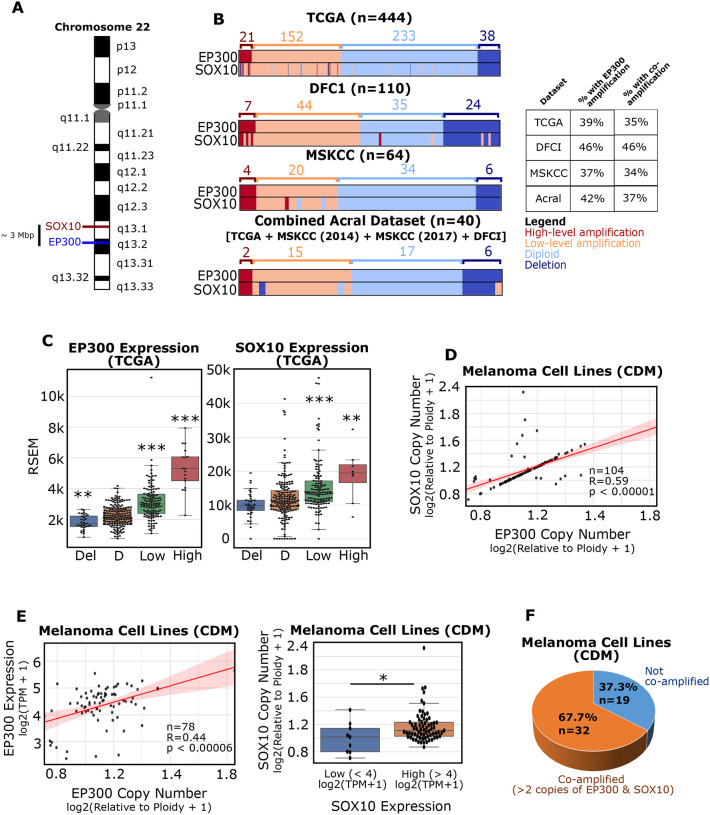
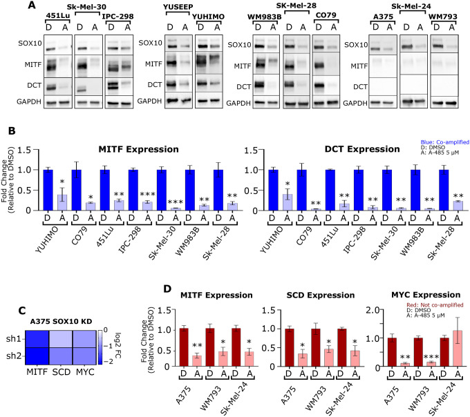
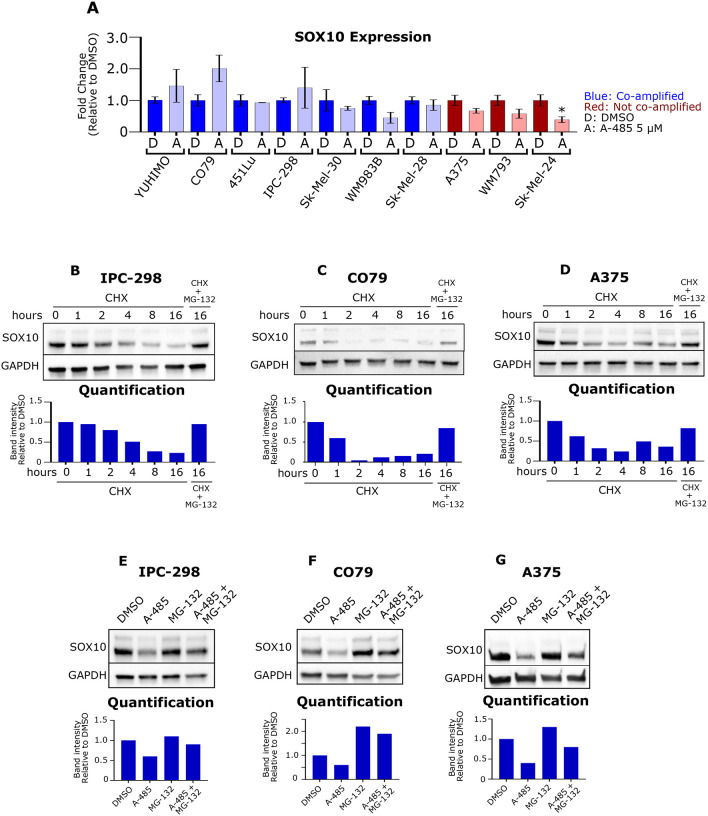
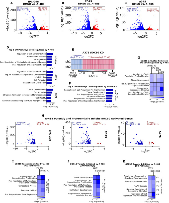
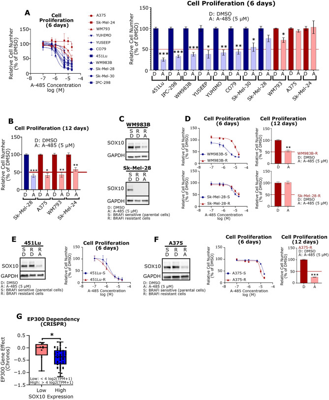
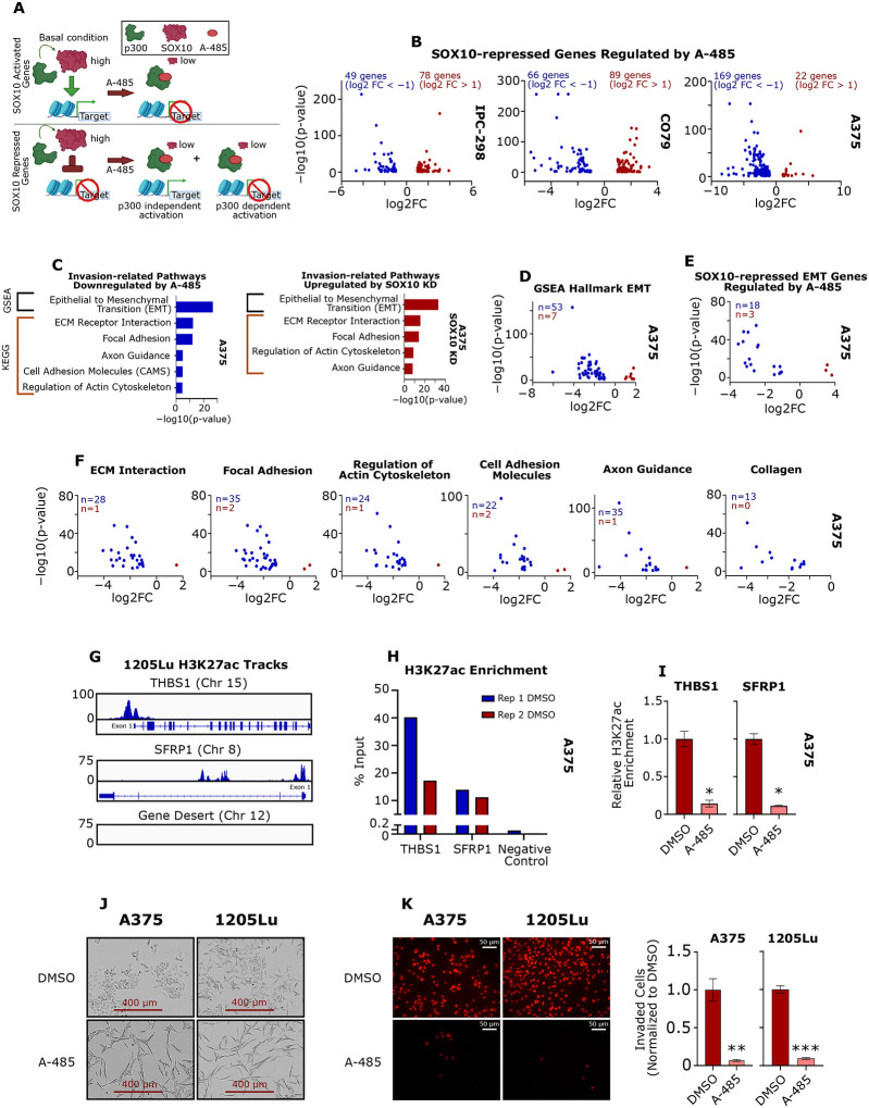
SOX10 is a lineage-specific transcription factor critical for melanoma tumor growth, while SOX10 loss-of-function drives the emergence of therapy-resistant, invasive melanoma phenotypes. A major challenge has been developing therapeutic strategies targeting SOX10's role in melanoma proliferation, while preventing a concomitant increase in tumor cell invasion. Here, we report that the lysine acetyltransferase (KAT) EP300 and SOX10 gene loci on Chromosome 22 are frequently co-amplified in melanomas, including UV-associated and acral tumors. We further show that p300 KAT activity mediates SOX10 protein stability and that the p300 inhibitor, A-485, downregulates SOX10 protein levels in melanoma cells via proteasome-mediated degradation. Additionally, A-485 potently inhibits proliferation of SOX10+ melanoma cells while decreasing invasion in AXLhigh/MITFlow melanoma cells through downregulation of metastasis-related genes. We conclude that the SOX10/p300 axis is critical to melanoma growth and invasion, and that inhibition of p300 KAT activity through A-485 may be a worthwhile therapeutic approach for SOX10-reliant tumors.

Keywords: A-485; SOX10; epigenetic; gene; melanoma; p300.


Conflict of interest statement

Conflict of Interest Statement P.A.C. is a co-founder of Acylin Therapeutics and a consultant for Abbvie regarding p300 acetyltransferase inhibitors. He also is a co-inventor on U.S. patent 11,565,994 B2 that concerns LSD1 and CoREST complex inhibitors. R.M.A. is a co-founder of Acylin Therapeutics.

Figures

Figure 1:
Figure 1:. EP300 is commonly co-amplified with SOX10 in melanomas.
(A) The EP300 and SOX10 genes are located in close proximity on Chromosome 22. (B) EP300 and SOX10 amplification prevalence in different melanoma datasets (TCGA, DFCI, MSKCC). Acral patients from multiple datasets were combined (Combined Acral). Amplification levels were defined by GISTIC2.0. (C) EP300 (left) and SOX10 (right) expression were analyzed for each copy number level, as defined by GISTIC2.0 (data from TCGA; Del: deletion; D: diploid; Low: low-level amplification; High: high-level amplification). (D) EP300 and SOX10 copy number are positively correlated in melanoma cell lines (data from Cancer Dependency Map [CDM]). (E) EP300 copy number and expression are positively correlated in melanoma cell lines (left). SOX10 copy number and expression are positively correlated in melanoma cell lines (right) (data CDM). (F) EP300/SOX10 co-amplifications (> 2 copies of EP300 and SOX10) are common in melanoma cell lines (data from CDM). *p < 0.05; **p < 0.005; ***p < 0.0005.
Figure 2:
Figure 2:. SOX10 signaling requires p300 KAT activity.
(A) A panel of melanoma cell lines were treated with DMSO (D) or 5 μM A-485 (A) for 24 hours. SOX10, MITF and DCT protein levels were assessed via immunoblotting. (B) A panel of EP300/SOX10 co-amplified melanoma cell lines were treated with DMSO (D) or 5 μM A-485 (A) for 24 hours. MITF and DCT expression were assessed via RT-qPCR. (C) MITF, SCD and MYC expression are downregulated by SOX10 knockdown (KD) in A375 cells. (D) A panel of melanoma cell lines without EP300/SOX10 co-amplifications were treated with DMSO (D) or 5 μM A-485 (A) for 24 hours. MITF, SCD and MYC expression were assessed via RT-qPCR. *p < 0.05; **p < 0.005; ***p < 0.0005.
Figure 3:
Figure 3:. A-485 induces proteasomal degradation of SOX10.
(A) A panel of melanoma cell lines were treated with DMSO (D) or 5 μM A-485 (A) for 24 hours. SOX10 expression was assessed via RT-qPCR. (B-D) IPC-298, CO79 and A375 cells were treated with 50 μg/mL cyclohexamide and/or 5 μM MG-132 for the indicated time. SOX10 protein levels were assessed via immunoblotting. (E-G) IPC-298, CO79 and A375 cells were treated with DMSO, 5 μM A-485, 5 μM MG-132, or their combination for 16 hours. SOX10 protein levels were assessed via immunoblotting. *p < 0.05.
Figure 4:
Figure 4:. A-485 treatment potently downregulates SOX10 target genes in melanoma cells.
(A-C) IPC-298, CO79 and A375 cells were treated with 5 μM A-485 for 24 hours and RNA-sequencing was performed. Volcano plots of differentially expressed genes in each cell line are depicted (DEG defined as FC > |2| and p < 0.05). (D) The top five Gene Ontology Biological Process pathways are shown for downregulated genes in each cell line. (E) A heatmap of DEGs resulting from SOX10 KD in A375 cells is shown (data from GSE50535). (F) The top five Gene Ontology Biological Process pathways are shown for SOX10-activated genes (i.e. genes downregulated by SOX10 KD). (G) A-485 downregulates Gene Ontology Biological Process pathways that are activated by SOX10 in IPC-298, CO79 and A375 cells. (H) Volcano plots of SOX10-activated genes differentially expressed due to A-485 treatment for IPC-298, CO79 and A375 cells. (I-K) Gene Ontology Biological Process pathways are shown for SOX10-activated genes that are downregulated by A-485.
Figure 5:
Figure 5:. A-485 potently inhibits proliferation of SOX10+ melanoma cell lines.
(A) A panel of melanoma cell lines were treated with A-485 (in a range of doses) for 6 days and their relative proliferation versus control was assessed (left). Results for 5 μM A-485 versus control are shown (right). (B) Cells that did not respond to A-485 within 6 days were pre-treated with 5 μM A-485 for 9 days and re-seeded for a 3-day proliferation assay and their relative proliferation versus control was assessed (12 days total of drug treatment). (C) WM983B-R and SK-Mel-28-R BRAF inhibitor resistant cells have low expression of SOX10 in comparison to parental cells. (D) The proliferation effects of short-term (6-day) or long-term (12-day) A-485 treatment of WM983B-R and SK-Mel-28-R cells was assessed. (E) 451Lu-R cells maintain SOX10 expression versus BRAFi-sensitive parental cells (left). 451Lu-S and 451Lu-R were treated with A-485 (in a range of doses) for 6 days and their relative proliferation versus control was assessed (right). (F) A375-R cells maintain SOX10 expression versus BRAFi-sensitive parental cells (left). A375-S and A375-R were treated with A-485 (in a range of doses) for 6 days and their relative proliferation versus control was assessed (middle). A375-S and A375-R cells were pre-treated 5 μM A-485 for 9 days and re-seeded for a 3-day proliferation assay and their relative proliferation versus control was assessed (right). (G) EP300 dependency scores are plotted for melanoma cell lines with low SOX10 (<4 log2(TPM+1) versus high SOX10 (>4 log2(TPM+1) expression in the CDM 22Q2 Public+Score, Chronos dataset. *p < 0.05; **p < 0.005; ***p < 0.0005.
Figure 6:
Figure 6:. p300 KAT activity is essential for activation of SOX10-repressed EMT markers.
(A) Diagram depicting the hypothesis that p300 activity is essential for expression of SOX10-activated genes, but SOX10-repressed genes may also require p300 for their activation. (B) Volcano plots of SOX10-repressed genes differentially expressed due to A-485 treatment for IPC-298, CO79 and A375 cells (DEG defined as FC > |2| and p < 0.05). (C) SOX10-repressed genes are enriched for genes involved in the epithelial-to-mesenchymal transition (EMT) and similar invasion-related pathways are downregulated by A-485. (D) Volcano plot of EMT genes differentially expressed due to A-485 treatment for A375 cells. (E) Volcano plot of SOX10-regulated EMT genes differentially expressed due to A-485 treatment for A375 cells. (F) Volcano plots of genes differentially expressed due to A-485 are shown for various KEGG invasion-related pathways identified in (C). Differentially expressed collagen genes are also shown. (G) H3K27ac is enriched at the promoters of EMT genes THBS1 and SFRP1, in comparison to a gene desert in invasive 1205Lu cells via ChIP-seq. (H) H3K27ac was confirmed to be enriched at the promoters of THBS1 and SFRP1, in comparison to a gene desert in A375 cells via ChIP-qPCR. (I) A-485 decreases H3K27ac enrichment at the promoters of THBS1 and SFRP1 as assessed by ChIP-qPCR. (J) 5 μM A-485 alters the cell morphology of A375 and 1205Lu cells after long-term (12-day) treatment. (K) A-485 potently inhibits invasion in A375 and 1205 after long-term (12-day) treatment. Representative images of invaded cells are shown on the left and quantification of invaded cells is shown on the right. *p < 0.05; **p < 0.005; ***p < 0.0005.

Similar articles

References

    1. Guo W., Wang H., and Li C. (2021). Signal pathways of melanoma and targeted therapy. Signal Transduct Target Ther 6, 424. 10.1038/s41392-021-00827-6. - DOI - PMC - PubMed
    1. Tang Y., Durand S., Dalle S., and Caramel J. (2020). EMT-Inducing Transcription Factors, Drivers of Melanoma Phenotype Switching, and Resistance to Treatment. Cancers (Basel) 12. 10.3390/cancers12082154. - DOI - PMC - PubMed
    1. Najem A., Soumoy L., Sabbah M., Krayem M., Awada A., Journe F., and Ghanem G.E. (2022). Understanding Molecular Mechanisms of Phenotype Switching and Crosstalk with TME to Reveal New Vulnerabilities of Melanoma. Cells 11. 10.3390/cells11071157. - DOI - PMC - PubMed
    1. Tsoi J., Robert L., Paraiso K., Galvan C., Sheu K.M., Lay J., Wong D.J.L., Atefi M., Shirazi R., Wang X., et al. (2018). Multi-stage Differentiation Defines Melanoma Subtypes with Differential Vulnerability to Drug-Induced Iron-Dependent Oxidative Stress. Cancer Cell 33, 890–904 e895. 10.1016/j.ccell.2018.03.017. - DOI - PMC - PubMed
    1. Shakhova O., Zingg D., Schaefer S.M., Hari L., Civenni G., Blunschi J., Claudinot S., Okoniewski M., Beermann F., Mihic-Probst D., et al. (2012). Sox10 promotes the formation and maintenance of giant congenital naevi and melanoma. Nat Cell Biol 14, 882–890. 10.1038/ncb2535. - DOI - PubMed

Publication types