[Preprint]. 2024 Jan 14:2024.01.13.575509.
doi: 10.1101/2024.01.13.575509.

The PCM scaffold enables RNA localization to centrosomes

Affiliations

The PCM scaffold enables RNA localization to centrosomes

Junnan Fang et al. bioRxiv. .

Update in

Abstract

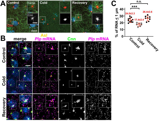
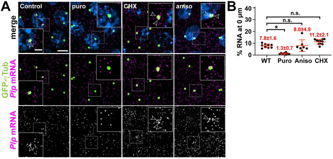
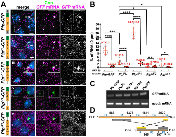
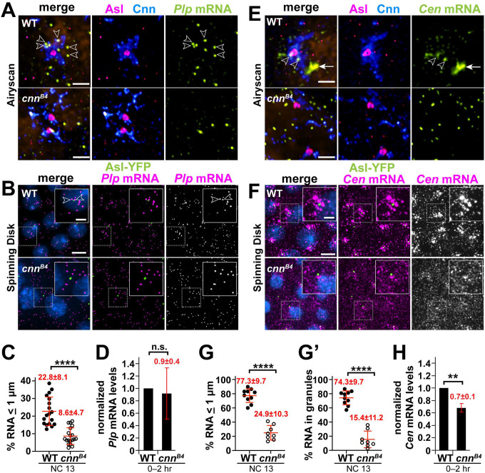
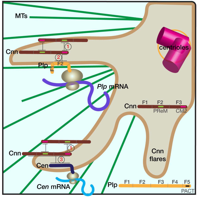
As microtubule-organizing centers, centrosomes direct assembly of the bipolar mitotic spindle required for chromosome segregation and genome stability. Centrosome activity requires the dynamic assembly of pericentriolar material (PCM), the composition and organization of which changes throughout the cell cycle. Recent studies highlight the conserved localization of several mRNAs encoded from centrosome-associated genes enriched at centrosomes, including Pericentrin-like protein (Plp) mRNA. However, relatively little is known about how RNAs localize to centrosomes and influence centrosome function. Here, we examine mechanisms underlying the subcellular localization of Plp mRNA. We find that Plp mRNA localization is puromycin-sensitive, and the Plp coding sequence is both necessary and sufficient for RNA localization, consistent with a co-translational transport mechanism. We identify regions within the Plp coding sequence that regulate Plp mRNA localization. Finally, we show that protein-protein interactions critical for elaboration of the PCM scaffold permit RNA localization to centrosomes. Taken together, these findings inform the mechanistic basis of Plp mRNA localization and lend insight into the oscillatory enrichment of RNA at centrosomes.

Keywords: Pericentrin; RNA localization; centrosome; co-translational transport.


Conflict of interest statement

Competing interest statement The authors have no competing interests to declare.

Figures

Figure 1.
Figure 1.. Plp mRNA localization requires microtubules.
Maximum intensity projections of (A) NC 11 embryos from the indicated conditions labeled with anti-Cnn (PCM; red) and α-Tub antibodies (microtubules; green), and DAPI (DNA; blue). (B) NC 12 P/p-GFP embryos from control, cold-treated, or recovery conditions labeled with GFP smFISH probes to show Plp mRNA distributions (magenta) and labelled with Cnn (green) and Asl (centrioles; yellow) antibodies and DAPI (blue). Dashed box regions are enlarged in insets. Arrowheads show Plp mRNA enrichments at centrosomes. (C) Quantification of GFP mRNA localization (within 1 μm from Asl). Each dot represents a measurement from a single embryo; see Table S1 for N embryos and RNA objects examined. Mean ± S.D. are displayed, n.s. not significant; ***p<0.001 by one-way ANOVA followed by Dunnett’s T3 multiple comparisons test. Scale bars: 5 μm; 2 μm (insets).
Figure 2.
Figure 2.. Plp mRNA localization to centrosomes is puromycin-sensitive.
(A) Maximum intensity projections of NC 13 embryos expressing GFP-γTub (green) labeled with Plp smFISH probes (magenta) and DAPI (blue) in controls or following treatment with translation inhibitors: puromycin (puro), cycloheximide (CHX), or anisomycin (aniso). Dashed box regions mark insets. Arrowheads show Plp mRNA enrichments at centrosomes. (B) Percentage of Plp mRNA localizing within 0 μm from the γTub surface. Mean ± S.D. are displayed, n.s. not significant; *p<0.05 by one-way ANOVA followed by Dunnett’s T3 multiple comparisons test. Scale bars: 5 μm; 2 μm (insets).
Figure 3.
Figure 3.. Plp mRNA localization requires sequences within the Plp CDS.
(A) Maximum intensity projections of NC 11 embryos expressing mCherry-Cnn (green) and labeled with GFP smFISH probes (magenta) to mark transgenic Plp mRNA localization and DAPI (blue) in the following genotypes: (i) Plp-GFP, (ii) UAS-PlpFL-GFP, (iii) UAS-PlpΔF1-GFP, (iv) UAS-PlpΔF2-GFP, and (v) UAS-PlpΔF5-GFP. Transgenes in (ii-v) were expressed using matGAL4 in the presence of endogenous Plp. Construct schematics are shown to the left. Arrowheads show RNA enrichments at centrosomes. (B) Quantification of GFP mRNA localization (0 |_im from Cnn surface). Each dot represents a measurement from a single embryo; see Table S1 for N embryos and RNA objects examined. The RNA channel was rotated 90° (+) and images re-quantified to assay the specificity of localization. (C) RT-PCR was used to assay the relative expression of the indicated GFP-tagged constructs from 0–2 hr embryos. (D) Schematic adapted from (Lerit et al., 2015) showing the two direct interaction modules between Plp and Cnn. Asterisk denotes the single point mutation (R1141H) that defines the cnn84 allele and abolishes the direct interaction between Plp F2 and Cnn CM2 (green bar). Mean ± S.D. are displayed, n.s. not significant; *p<0.05; ***p<0.001; ****p<0.0001 by one-way ANOVA followed by Dunnett’s T3 multiple comparisons test. Uncropped gels are available at <10.6084/m9.figshare.24926298>. Scale bars: 5 μm; 2 μm (insets).
Figure 4.
Figure 4.. The centrosome scaffold permits mRNA localization.
Maximum intensity projections of NC 13 control and cnnB4 embryos labeled with (A and B) Pip mRNA or (E and F) Cen mRNA smFISH probes. In (A and E), embryos were co-stained with smFISH probes (green), anti-Cnn (blue) and Asl (magenta) antibodies, and DAPI (orange; nuclei), then imaged using a Zeiss LSM 880 Airyscan. For (B and F), embryos expressing Asl-YFP (green) were labeled with smFISH probes (magenta) and DAPI (blue) then imaged by spinning disk confocal microscopy. (C) Quantification shows the percentage of Pip mRNA localizing within 1 μm from the Asl surface. (D) Levels of Pip mRNA or (H) Cen mRNA were normalized to RP49 as detected by qPCR from 0–2 hr WT versus cnn84 embryos and displayed relative to the WT control. (G) The percentage of Cen mRNA localizing and (G’) residing within granules (defined as > 4 RNA molecules per object [17]) within 1 μm from the Asl surface. Each dot represents a measurement from a single embryo; see Table S1 for N embryos and RNA objects examined. Mean ± S.D. are displayed, n.s., not significant or ****p<0.0001 by unpaired student t-test. Scale bars: (A and E) 1 μm; (B and F) 5 μm; 2 μm (insets).
Figure 5.
Figure 5.. The centrosome scaffold supports Cen mRNA localization and granule formation.
Maximum intensity projections of NC 13 (A) WT or (B) PlpGLC embryos labeled with Cen smFISH probes (magenta), Asl antibodies (green), and DAPI (blue). Charts show the percentage of Cen mRNA (B) localizing or (B’) residing within granules (> 4 RNA molecules per object) within 1 pm from the Asl surface. Each dot represents a measurement from a single embryo; see Table S1 for N embryos and RNA objects examined. (C) Levels of Cen mRNA were normalized to RP49 mRNA as detected by qPCR from 0–2 hr WT versus PlpGLC embryos and displayed relative to the WT control. Mean ± S.D. are displayed, n.s. not significant, and ****p<0.0001 by unpaired student t-test. Scale bars: 5 μm; 2 μm (insets).
Figure 6.
Figure 6.. Schematic of RNA localization to centrosomes.
The cartoon shows part of a centrosome with extended Cnn flares (brown), which contribute to PCM scaffolding. Elaboration of the PCM scaffold requires oligomerization of Cnn between its PReM and CM2 motifs (interaction 1), and a direct interaction between CM2 and PLP F2 (interaction 2; [61, 39, 24]). Simplified protein architectures of Cnn and Plp are noted in the figure. We propose that the Plp F2-Cnn CM2 interaction helps transmit and/or anchor the Plp mRNA-protein complex to the centrosome. Accordingly, microtubules (MTs, green), are required both for the extension of Cnn flares and the localization of Plp mRNA to centrosomes [24, 46, 39, 62]. Cen mRNA also localizes to the centrosome via co-translational transport, and Cen protein interacts directly with Cnn (interaction 3). Mutant analysis indicates that an intact PCM scaffold is required for the localization of both Plp and Cen mRNAs. We further propose that the temporal control of PCM scaffold elaboration (i.e., extension of Cnn flares) similarly regulates RNA localization to centrosomes.

References

    1. Nigg E. A. and Raff J. W., “Centrioles, Centrosomes, and Cilia in Health and Disease,” Cell, 2009. 139 (4): 663–678. - PubMed
    1. Doxsey S., Zimmerman W., and Mikule K., “Centrosome control of the cell cycle,” Trends in Cell Biology, 2005. 15 (6): 303–311. - PubMed
    1. Vertii A., Hehnly H., and Doxsey S., “The Centrosome, a Multitalented Renaissance Organelle,” Cold Spring Harb Perspect Biol, 2016. 8 (12): a025049. - PMC - PubMed
    1. Müller H. et al. , “Proteomic and functional analysis of the mitotic Drosophila centrosome,” The EMBO journal, 2010. 29 (19): 3344–3357. - PMC - PubMed
    1. Gupta G. D. et al. , “A Dynamic Protein Interaction Landscape of the Human Centrosome-Cilium Interface,” Cell, 2015. 163 (6): 1484–99. - PMC - PubMed

Publication types