

Delineating thrombus versus myxoma: Perioperative 3D transesophageal echocardiography to the rescue!
- PMID: 38469175
- PMCID: PMC10927041
- DOI: 10.25259/JCIS_136_2023
Delineating thrombus versus myxoma: Perioperative 3D transesophageal echocardiography to the rescue!
Abstract
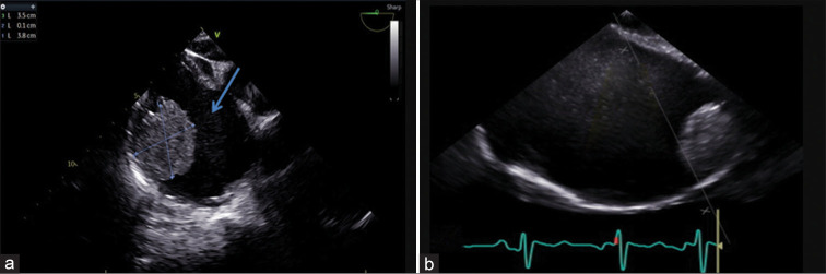
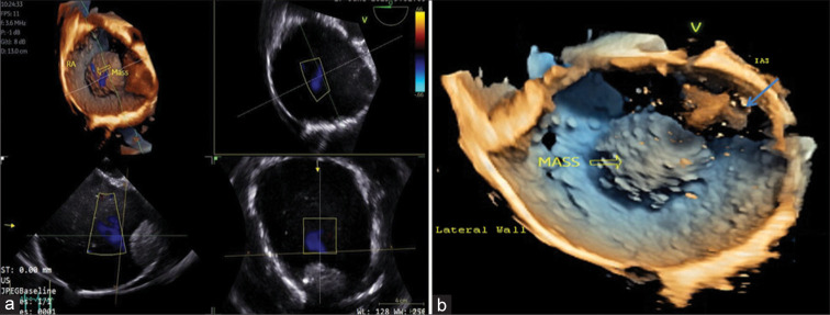
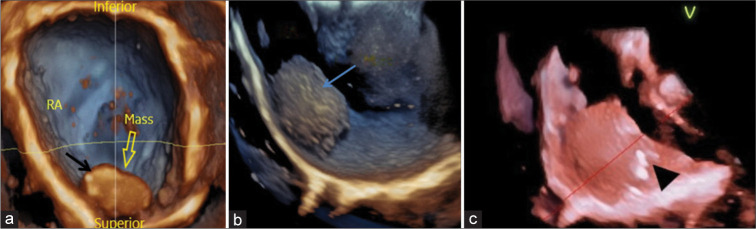
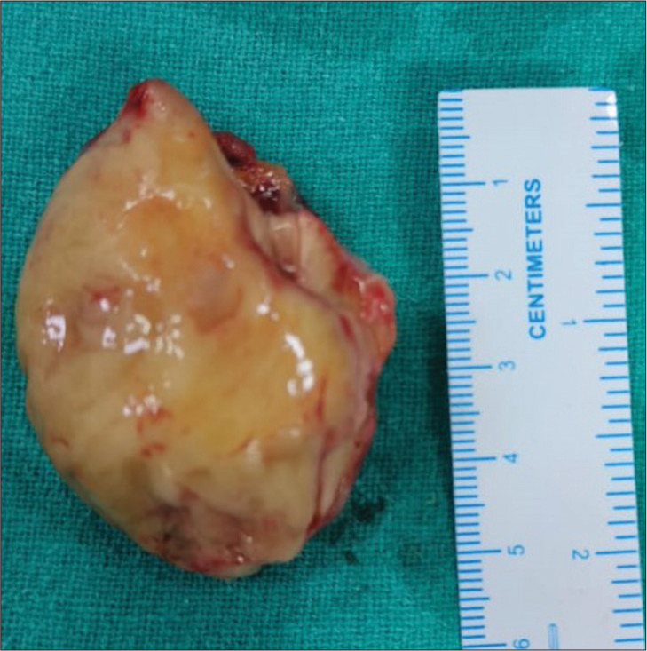
Cardiac masses are a significant cause of patient morbidity and mortality by virtue of their symptoms and surgical removal. Preoperative diagnosis of a cardiac mass is usually based on clinical correlation and transthoracic echocardiography findings. Myxomas are the most common benign cardiac tumors, commonly occurring in the left atrium attached to the interatrial septum near the fossa ovalis. Although, at times atypical location and unusual morphology may pose a diagnostic dilemma with 2D echocardiography. 3D echocardiography with its multifaceted advantages, including multiplanar cropping abilities and superior imaging quality can help distinguish between a clot and a myxoma.
Keywords: 3D echocardiography; Myxoma; Thrombus.
© 2024 Published by Scientific Scholar on behalf of Journal of Clinical Imaging Science.
Conflict of interest statement
There are no conflicts of interest.
Figures
Similar articles
-
[Integrated echocardiography (transthoracic-transesophageal) in the differential diagnosis of left atrial myxoma. Description of three clinical cases].Cardiologia. 1998 May;43(5):515-8. Cardiologia. 1998. PMID: 9701883 Italian.
-
[Advantages of transesophageal color Doppler echocardiography in the diagnosis and surgical treatment of cardiac masses].J Cardiol. 1990;20(3):701-14. J Cardiol. 1990. PMID: 2131359 Japanese.
-
Left atrial cardiac myxoma. Two unusual cases studied by 3D echocardiography.BMJ Case Rep. 2014 Nov 17;2014:bcr2014205938. doi: 10.1136/bcr-2014-205938. BMJ Case Rep. 2014. PMID: 25404247 Free PMC article.
-
Atrial myxoma: case report and a review of the literature.Heart Dis. 2003 May-Jun;5(3):224-30. doi: 10.1097/01.hdx.0000074515.95567.92. Heart Dis. 2003. PMID: 12783636 Review.
-
[Contribution of transesophageal echocardiography in the diagnosis of intra- and para-cardiac masses].Arch Mal Coeur Vaiss. 1993 Mar;86(3):331-8. Arch Mal Coeur Vaiss. 1993. PMID: 8215768 Review. French.
References
Publication types
LinkOut - more resources
Full Text Sources