

Microwave-enhanced antibacterial activity of polydopamine-silver hybrid nanoparticles
- PMID: 38469191
- PMCID: PMC10926840
- DOI: 10.1039/d3ra07543e
Microwave-enhanced antibacterial activity of polydopamine-silver hybrid nanoparticles
Abstract
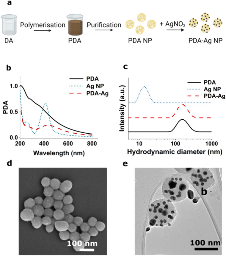
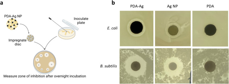
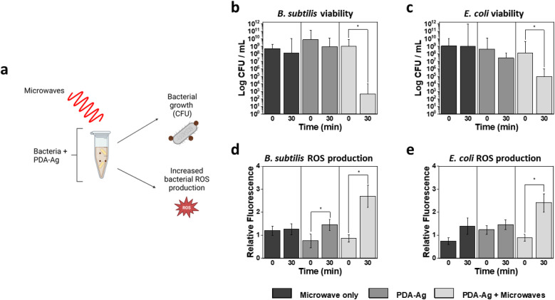
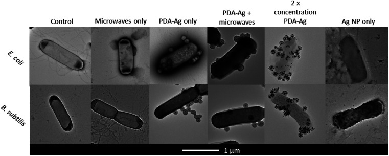
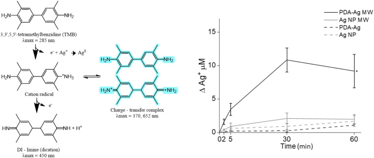
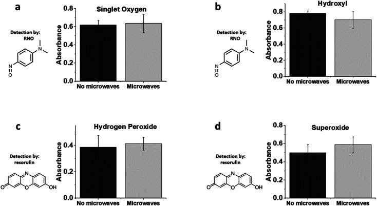
The ever-increasing risks posed by antibiotic-resistant bacteria have stimulated considerable interest in the development of novel antimicrobial strategies, including the use of nanomaterials that can be activated on demand and result in irreversible damage to pathogens. Microwave electric field-assisted bactericidal effects on representative Gram-negative and Gram-positive bacterial strains were achieved in the presence of hybrid polydopamine-silver nanoparticles (PDA-Ag NPs) under low-power microwave irradiation using a resonant cavity (1.3 W, 2.45 GHz). A 3-log reduction in the viability of bacterial populations was observed within 30 minutes which was attributed to the attachment of PDA-Ag NPs and associated membrane disruption in conjunction with the production of intra-bacterial reactive oxygen species (ROS). A synergistic effect between PDA and Ag has been demonstrated whereby PDA acts both as an Ag NP carrier and a microwave enhancer. These properties together with the remarkable adhesivity of PDA are opening a route to design of antibacterial adhesives and surface coatings for prevention of biofilm formation.
This journal is © The Royal Society of Chemistry.
Conflict of interest statement
There are no conflicts to declare.
Figures
Similar articles
-
Engineering of Near-Infrared-Activated Lignin-Polydopamine-Nanosilver Composites for Highly Efficient Sterilization.ACS Appl Bio Mater. 2022 Aug 15. doi: 10.1021/acsabm.2c00474. Online ahead of print. ACS Appl Bio Mater. 2022. PMID: 35969409
-
Sonochemically-Produced Metal-Containing Polydopamine Nanoparticles and Their Antibacterial and Antibiofilm Activity.Langmuir. 2016 May 24;32(20):5201-12. doi: 10.1021/acs.langmuir.6b00576. Epub 2016 May 11. Langmuir. 2016. PMID: 27133213
-
Pyroelectric Janus nanomotors for synergistic electrodynamic-photothermal-antibiotic therapies of bacterial infections.Acta Biomater. 2023 May;162:20-31. doi: 10.1016/j.actbio.2023.03.012. Epub 2023 Mar 16. Acta Biomater. 2023. PMID: 36931421
-
Antibacterial activities of transient metals nanoparticles and membranous mechanisms of action.World J Microbiol Biotechnol. 2019 Oct 14;35(10):162. doi: 10.1007/s11274-019-2742-6. World J Microbiol Biotechnol. 2019. PMID: 31612285 Review.
-
Nanotechnology as a therapeutic tool to combat microbial resistance.Adv Drug Deliv Rev. 2013 Nov;65(13-14):1803-15. doi: 10.1016/j.addr.2013.07.011. Epub 2013 Jul 24. Adv Drug Deliv Rev. 2013. PMID: 23892192 Review.
Cited by
-
Bioinspired Nanoplatforms: Polydopamine and Exosomes for Targeted Antimicrobial Therapy.Polymers (Basel). 2025 Jun 16;17(12):1670. doi: 10.3390/polym17121670. Polymers (Basel). 2025. PMID: 40574198 Free PMC article. Review.
-
Evaluation of enamel remineralization potential and anticariogenic efficacy of polydopamine coated biogenic amorphous calcium phosphate.Clin Oral Investig. 2025 May 20;29(6):302. doi: 10.1007/s00784-025-06384-4. Clin Oral Investig. 2025. PMID: 40389610
-
Biomineralization of dental tissues with natural drugs: a comprehensive review.Saudi Dent J. 2025 Jul 15;37(4-6):29. doi: 10.1007/s44445-025-00036-9. Saudi Dent J. 2025. PMID: 40663199 Free PMC article. Review.
References
-
- O'Neill J., Tackling drug-resistant infections globally: final report and recommendations, Wellcome Trust commissioned Review on Antimicrobial Resistance, 2016, https://amr-review.org/home.html
LinkOut - more resources
Full Text Sources
Miscellaneous