

Nanofibrous electrospun scaffold doped with hydroxyapatite derived from sand lobster shell (Panulirus homarus) for bone tissue engineering
- PMID: 38469192
- PMCID: PMC10925909
- DOI: 10.1039/d4ra00619d
Nanofibrous electrospun scaffold doped with hydroxyapatite derived from sand lobster shell (Panulirus homarus) for bone tissue engineering
Abstract
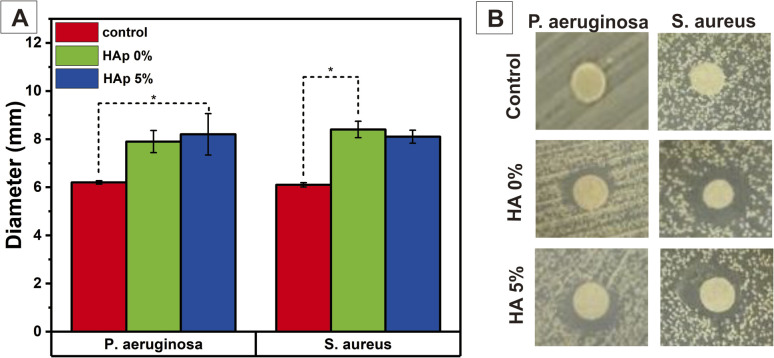
Healing of significant segmental bone defects remains a challenge, and various studies attempt to make materials that mimic bone structures and have biocompatibility, bioactivity, biodegradability, and osteoconductivity to native bone tissues. In this work, a nanofiber scaffold membrane of polyvinyl alcohol (PVA)/polyvinylpyrrolidone (PVP)/chitosan (CS) combined with hydroxyapatite (HAp) from sand lobster (SL; Panulirus homarus) shells, as a calcium source, was successfully synthesized to mimic the nanoscale extracellular matrix (ECM) in the native bone. The HAp from SL shells was synthesized by co-precipitation method with Ca/P of 1.67 and incorporated into the nanofiber membrane PVA/PVP/CS synthesized by the electrospinning method with varying concentrations, i.e. 0, 1, 3, and 5% (w/v). Based on the morphological and physicochemical analysis, the addition of HAp into the nanofiber successfully showed incorporation into the nanofiber with small agglomeration at HAp concentrations of 1, 3, and 5% (w/v). This led to a smaller fiber diameter with higher concentration of Hap, and incorporating HAp into the nanofiber could improve the mechanical properties of the nanofiber closer to the trabecula bone. Moreover, in general, swelling due to water absorption increases due to higher hydrophilicity at higher HAp concentrations and leads to the improvement of the degradation process and protein adsorption of the nanofiber. Biomineralization in a simulated body fluid (SBF) solution confirms that the HAp in the nanofiber increases bioactivity, and it can be seen that more apatite is formed during longer immersion in the SBF solution. The nanofiber PVA/PVP/CS HAp 5% has the most potential for osteoblast (MC3T3E1) cell viability after being incubated for 24 h, and it allowed the cell to attach and proliferate. Additionally, the higher HAp concentration in the nanofiber scaffold membrane can significantly promote the osteogenic differentiation of MC3T3E1 cells. Overall, the PVA/PVP/CS/HAp 5% nanofiber scaffold membrane has the most potential for bone tissue engineering.
This journal is © The Royal Society of Chemistry.
Conflict of interest statement
The authors declare that they have no known competing financial interests or personal relationships that could influence the work in this article.
Figures


Similar articles
-
Porous scaffold hydroxyapatite from sand lobster shells (Panulirus homarus) using polyethylene oxide/chitosan as polymeric porogen for bone tissue engineering.J Biomed Mater Res B Appl Biomater. 2024 Jan;112(1):e35341. doi: 10.1002/jbm.b.35341. Epub 2023 Oct 25. J Biomed Mater Res B Appl Biomater. 2024. PMID: 37877433
-
Electrospun polyvinyl alcohol nanofiber scaffolds incorporated strontium-substituted hydroxyapatite from sand lobster shells: synthesis, characterization, and in vitro biological properties.Biomed Mater. 2024 Sep 23. doi: 10.1088/1748-605X/ad7e92. Online ahead of print. Biomed Mater. 2024. PMID: 39312949
-
Nanofibrous poly(vinyl alcohol)/chitosan contained carbonated hydroxyapatite nanoparticles scaffold for bone tissue engineering.Mater Sci Eng C Mater Biol Appl. 2020 Feb;107:110347. doi: 10.1016/j.msec.2019.110347. Epub 2019 Oct 23. Mater Sci Eng C Mater Biol Appl. 2020. PMID: 31761152
-
New Prospects in Nano Phased Co-substituted Hydroxyapatite Enrolled in Polymeric Nanofiber Mats for Bone Tissue Engineering Applications.Ann Biomed Eng. 2021 Sep;49(9):2006-2029. doi: 10.1007/s10439-021-02810-2. Epub 2021 Aug 10. Ann Biomed Eng. 2021. PMID: 34378121 Review.
-
The efficiency of PCL/HAp electrospun nanofibers in bone regeneration: a review.J Med Eng Technol. 2021 Oct;45(7):511-531. doi: 10.1080/03091902.2021.1893396. Epub 2021 Jul 12. J Med Eng Technol. 2021. PMID: 34251971 Review.
Cited by
-
A comparative X-ray diffraction analysis of Sr2+substituted hydroxyapatite from sand lobster shell waste using various methods.Heliyon. 2025 Jan 8;11(2):e41781. doi: 10.1016/j.heliyon.2025.e41781. eCollection 2025 Jan 30. Heliyon. 2025. PMID: 39877603 Free PMC article.
-
Dual-Layer Natamycin and Boric-Acid-Reinforced PVA/Chitosan by 3D Printing and Electrospinning Method: Characterization and In Vitro Evaluation.Polymers (Basel). 2025 Jun 17;17(12):1673. doi: 10.3390/polym17121673. Polymers (Basel). 2025. PMID: 40574201 Free PMC article.
-
Nano-Hydroxyapatite/Poly(methyl methacrylate) Composite Bone Scaffold: Surfactant Surface Effects.Polymers (Basel). 2025 Apr 23;17(9):1148. doi: 10.3390/polym17091148. Polymers (Basel). 2025. PMID: 40362932 Free PMC article.
-
Polysaccharide Hydroxyapatite (Nano)composites and Their Biomedical Applications: An Overview of Recent Years.ACS Omega. 2024 Jul 2;9(28):30035-30070. doi: 10.1021/acsomega.4c02170. eCollection 2024 Jul 16. ACS Omega. 2024. PMID: 39035931 Free PMC article. Review.
References
-
- Permatasari H. A. Yusuf Y. Characteristics of Carbonated Hydroxyapatite Based on Abalone Mussel Shells (Halioitis asinina) Synthesized by Precipitation Method with Aging Time Variations. IOP Conf. Ser.: Mater. Sci. Eng. 2019;546(4):042031.
-
- Stevens M. M. Biomaterials for bone tissue engineering. Mater. Today. 2008;11(5):18–25.
-
- Akram M. Ahmed R. Shakir I. Ibrahim W. A. W. Hussain R. Extracting hydroxyapatite and its precursors from natural resources. J. Mater. Sci. 2014;49(4):1461–1475.
LinkOut - more resources
Full Text Sources
Miscellaneous