. 2024 Feb 26:14:1323422.
doi: 10.3389/fonc.2024.1323422. eCollection 2024.

Oncopig bladder cancer cells recapitulate human bladder cancer treatment responses in vitro

Affiliations

Oncopig bladder cancer cells recapitulate human bladder cancer treatment responses in vitro

Natália V Segatto et al. Front Oncol. .

Abstract

Introduction: Bladder cancer is a common neoplasia of the urinary tract that holds the highest cost of lifelong treatment per patient, highlighting the need for a continuous search for new therapies for the disease. Current bladder cancer models are either imperfect in their ability to translate results to clinical practice (mouse models), or rare and not inducible (canine models). Swine models are an attractive alternative to model the disease due to their similarities with humans on several levels. The Oncopig Cancer Model has been shown to develop tumors that closely resemble human tumors. However, urothelial carcinoma has not yet been studied in this platform.

Methods: We aimed to develop novel Oncopig bladder cancer cell line (BCCL) and investigate whether these urothelial swine cells mimic human bladder cancer cell line (5637 and T24) treatment-responses to cisplatin, doxorubicin, and gemcitabine in vitro.

Results: Results demonstrated consistent treatment responses between Oncopig and human cells in most concentrations tested (p>0.05). Overall, Oncopig cells were more predictive of T24 than 5637 cell therapeutic responses. Microarray analysis also demonstrated similar alterations in expression of apoptotic (GADD45B and TP53INP1) and cytoskeleton-related genes (ZMYM6 and RND1) following gemcitabine exposure between 5637 (human) and Oncopig BCCL cells, indicating apoptosis may be triggered through similar signaling pathways. Molecular docking results indicated that swine and humans had similar Dg values between the chemotherapeutics and their target proteins.

Discussion: Taken together, these results suggest the Oncopig could be an attractive animal to model urothelial carcinoma due to similarities in in vitro therapeutic responses compared to human cells.

Keywords: Oncopig cancer model; bladder cancer; cisplatin; doxorubicin; gemcitabine; in silico; microarray.


Conflict of interest statement

LBS, LJ, CB, and KS work for Sus Clinicals, which provides the Oncopig and other pig-based preclinical testing services to customers. The remaining authors declare that the research was conducted in the absence of any commercial or financial relationships that could be construed as a potential conflict of interest. The author(s) declared that they were an editorial board member of Frontiers, at the time of submission. This had no impact on the peer review process and the final decision.

Figures

Figure 1
Figure 1
Human and Oncopig cell line sensitivity to chemotherapeutics. Human bladder cancer cell lines (5637 and T24) and Oncopig BCCLs treated with commercial chemotherapeutics cisplatin, gemcitabine, or doxorubicin. (A) Antiproliferative activity in cells treated for 48 and 72h obtained by MTT assay. Y axis: Cell viability (relative to % of each cell line control). (B) Cytotoxicity results obtained by Live/Dead assay. Y-axis: number of viable cells (% of control). (C) ViaCount assay results. Y-axis: number of viable cells/mL (% of control). All data are expressed as mean ± SEM of three independent experiments. Asterisks (*) indicate significant differences between Oncopig BCCL and 5637 means and hashtag (#) indicates significant differences between Oncopig BCCL and T24 means at the same drug concentration. * or #= p<0.05. **= p<0.01. ***= p<0.001. The Y axis dotted line represents 50% of growth inhibition.
Figure 2
Figure 2
Apoptosis induction in Oncopig and human cell lines after drug treatment. Human bladder cancer cell lines (5637 and T24) and Oncopig BCCLs treated with commercial chemotherapeutics cisplatin, gemcitabine, or doxorubicin for 48h and subjected to DAPI assay. (A) Apoptosis induction percentage. Asterisks (*) indicate significant differences between Oncopig BCCL and 5637 means and hashtag (#) indicates significant differences between Oncopig BCCL and T24 means at the same drug concentration. * or #= p<0.05. **= p<0.001. Y axis: Apoptosis induction percentage. X axis: drug treatment. (B) Confocal microscopy images. Blue represents cell nucleus staining (emission ~460 nm). White arrows indicate apoptotic cells that are distinguished by their higher fluorescence and condensed DNA.
Figure 3
Figure 3
Summary of apoptosis pathways in human and Oncopig cells. Summarized apoptotic pathways altered by gemcitabine treatment in Oncopig BCCL and 5637 cells evaluated by microarray. There were multiple possible cellular pathways involved in apoptosis induction, such as: TP53INP1/P53/BCL/BAX/CASPASE mitochondrial intrinsical pathway; TP53INP1/TP73 activation; FAS/RB1/GADD45B/CASPASE pathway and cell rounding up by RND1 and/or ZMYM6.
Figure 4
Figure 4
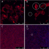
Cytoskeleton reorganization. Blue represents cells’ nucleus staining with DAPI (emission ~460 nm). Red represents cytoskeleton staining with Texas Red (emission ~615 nm). 200x magnification. White circles demonstrate examples of cell rounding. (A, B): Oncopig cells (BCCL). (C, D): human cells (5637). (A, C): untreated cells. (B, D): cells treated with 1µm of gemcitabine.

Similar articles

Cited by

References

    1. Bray F, Ferlay J, Soerjomataram I, Siegel RL, Torre LA, Jemal A. Global cancer statistics 2018: GLOBOCAN estimates of incidence and mortality worldwide for 36 cancers in 185 countries. CA Cancer J Clin (2018) 68(6):394–424. doi: 10.3322/caac.21492 - DOI - PubMed
    1. (ASCO) AS of CO . Bladder Cancer: Introduction (2020). Available at: https://www.cancer.net/cancer-types/bladder-cancer/introduction.
    1. Kamat AM, Flaig TW, Grossman HB, Konety B, Lamm D, O’Donnell MA, et al. . Expert consensus document: Consensus statement on best practice management regarding the use of intravesical immunotherapy with BCG for bladder cancer. Nat Rev Urol (2015) 12:225–35. doi: 10.1038/nrurol.2015.58 - DOI - PubMed
    1. Kamat AM, Hahn NM, Efstathiou JA, Lerner SP, Malmström PU, Choi W, et al. . Bladder cancer. Lancet (2016) 388(10061):2796–810. doi: 10.1016/S0140-6736(16)30512-8 - DOI - PubMed
    1. Barabas K, Milner R, Lurie D, Adin C. Cisplatin: A review of toxicities and therapeutic applications. Veterinary Comp Oncol (2008) 6:1–18. doi: 10.1111/j.1476-5829.2007.00142.x - DOI - PubMed